David F. Cuevas Cueto, MD · Updated September 27, 2026 · Sources: SCCT 2026 and CAD-RADS 2.0
CAD-RADS 2.0 summarizes the findings of coronary CT angiography in a single per-patient category, followed by plaque burden and modifiers. This section explains how each category is assigned, the order in which the code is written, and how CT-FFR or CT perfusion results are incorporated. The management summarized here applies to stable chest pain.
Key points
The CAD-RADS category is assigned once per patient and is defined by the clinically most relevant stenosis, which is almost always the highest-grade one.
CAD-RADS 0 requires no plaque and no stenosis, so plaque without stenosis is already enough for CAD-RADS 1.
Left main stenosis of 50 % or more is 4B even if no vessel reaches 70 %, as is three-vessel disease with 70 % or more.
If a segment is nonevaluable and an evaluable segment reaches 50 %, the stenosis is graded and /N is added. If none reaches 50 %, the category is N.
The code is written in the order category, P, N, HRP, I, S, G and E, separated by slashes, and P is not assigned in CAD-RADS 0.
With CT-FFR measured 1 to 2 cm distal to the lesion, 0.75 or less is I+, 0.76 to 0.80 is I±, and above 0.80 is I−.
CAD-RADS string reader
Type or paste a string as it appears in a report to see what each piece means, with a link to its term.
Examples:
The reader explains CAD-RADS 2.0 nomenclature; it does not interpret a specific study or recommend management.
Table 1. CAD-RADS 2.0 categories and suggested management in the patient with stable chest pain. These are consensus suggestions, to be interpreted together with the impression and the clinical context.
Category
Maximal stenosis
Interpretation
Suggested management (stable chest pain)
CAD-RADS 0
0 % (no plaque or stenosis)
No coronary artery disease
No further testing. Consider nonatherosclerotic causes.
CAD-RADS 1
1–24 %, or plaque without stenosis
Minimal nonobstructive disease
No further testing. Preventive therapy according to P, intensive with P3–P4.
CAD-RADS 2
25–49 %
Mild nonobstructive disease
No further testing. Preventive therapy according to P, intensive with P3–P4.
Consider invasive angiography, functional or viability assessment. Intensive prevention.
CAD-RADS N
Nondiagnostic study
Obstructive disease cannot be excluded
Additional or alternative evaluation may be needed.
Source: CAD-RADS 2.0, Table 4 (PMC full text, unpaginated); stenosis ranges also in SCCT 2026, p. 15. In the CAD-RADS 5 row, Table 4 reads «total coronary occlusion or sub-total occlusion»; the glossary keeps «total occlusion» because the category requires 100 %.
Also seen as:Coronary Artery Disease-Reporting and Data System, CAD-RADS score(Informal), CAD-RADS classification, CADRADS
CAD-RADS 2.0 assigns a single category to each patient. It is defined by the clinically most relevant stenosis, usually the highest-grade one, and only vessels larger than 1.5 mm are graded.
In practice. Each segment is graded first and its stenosis is recorded in the findings. The most severe stenosis defines the category, unless left main stenosis or three-vessel disease raises it to 4B. Lesser lesions are described in the findings.
Pitfall. Assigning a category to each vessel, such as «LAD CAD-RADS 3 and RCA CAD-RADS 2». The patient receives one category, and each lesion is described in the findings.
Other sources. SCOT-HEART classified each patient into three groups: normal coronary arteries, nonobstructive disease, and obstructive disease, defined as stenosis greater than 70 %, or greater than 50 % in the left main. CAD-RADS 2.0 assigns one of eight categories. (Williams 2019, SCOT-HEART, Methods)
On the map. The map takes the highest grade among the marked segments and shows a single category with its reason, for example «Moderate stenosis (50–69%)». The template describes each vessel in its own paragraph. Open the example on the map (CAD-RADS 3/P2) →
How the map's template words it
Left anterior descending (LAD): mid segment: partially calcified plaque resulting in moderate stenosis (50-69%); the remainder is unremarkable. Branches D1, D2: no plaque.
Circumflex (LCx): proximal segment: calcified plaque causing minimal stenosis (1-24%); the rest of the vessel is unremarkable. Branches OM1, OM2: no plaque.
Right coronary (RCA): proximal segment: calcified plaque causing mild stenosis (25-49%); the rest of the vessel is unremarkable. Branches AM, R-PDA, R-PLB: no plaque.
Source: CAD-RADS 2.0, Table 4, footnote · «The CAD-RADS classification should be applied on a per-patient basis for the clinically most relevant (usually highest-grade) stenosis.»
Also seen as:absence of CAD(CAD-RADS 2.0 interpretation of CAD-RADS 0), minimal non-obstructive CAD(CAD-RADS 2.0 interpretation of CAD-RADS 1)
A study is CAD-RADS 0 only if there is no plaque and no stenosis. Any plaque raises it to at least CAD-RADS 1, even if it does not narrow the lumen, such as plaque with positive remodeling and no stenosis.
In practice. Before calling a study normal, review each segment on cross-sectional views and multiplanar reconstructions for small non-calcified plaque. SCCT 2026 reserves the impression of a normal study for the absence of plaque and stenosis.
Pitfall. Calling a study CAD-RADS 0 when the Agatston score is 0 but non-calcified plaque is present. That plaque is enough for CAD-RADS 1, and plaque burden is assigned.
On the map. A plaque marked at 0 % stenosis is written as «plaque without significant stenosis» and raises the category to 1. In category 0 the map does not show P, HRP or I. Open the example on the map (CAD-RADS 1/P1) →
How the map's template words it
Left anterior descending (LAD): proximal segment: plaque without significant stenosis with positive remodeling as an isolated feature; the remainder is unremarkable. Branches D1, D2: no plaque.
Source: CAD-RADS 2.0, Table 4, footnote b · «CAD-RADS 1 - This category should also include the presence of plaque with positive remodeling and no evidence of stenosis.»
Also seen as:mild non-obstructive CAD(CAD-RADS 2.0 interpretation of CAD-RADS 2)
In CAD-RADS 2 the maximal stenosis is mild (25–49 %) and disease is nonobstructive. In CAD-RADS 3 it is moderate (50–69 %), and CAD-RADS 2.0 already suggests considering functional testing.
In practice. Stenosis is estimated in at least two perpendicular planes, comparing the minimal luminal diameter with a normal reference segment. When the grade is close to 50 %, review the lesion on cross-sectional views before choosing between 2 and 3.
Pitfall. Reading CAD-RADS 3 as a direct indication for invasive angiography. In the stable patient, CAD-RADS 2.0 suggests considering functional testing.
Other sources. In invasive angiography, the 2021 ACC/AHA/SCAI revascularization guideline calls a 40 to 69 % stenosis intermediate, which generally warrants physiologic assessment. In CAD-RADS 2.0, a 40 to 49 % stenosis is CAD-RADS 2. (ACC/AHA/SCAI 2021 revascularization guideline, Section 4.1)
On the map. With the 2 and 3 buttons, the template writes the term and its range, such as «moderate stenosis (50-69%)». With the slider it writes the percentage, such as «approximately 55% stenosis». Open the example on the map (CAD-RADS 3/P1) →
How the map's template words it
Circumflex (LCx): proximal segment: non-calcified plaque causing approximately 55% stenosis; the rest of the vessel is unremarkable. Branches OM1, OM2: no plaque.
Right coronary (RCA): mid segment: partially calcified plaque producing mild stenosis (25-49%); the rest of the vessel is unremarkable. Branches AM, R-PDA, R-PLB: no plaque.
Source: CAD-RADS 2.0, Section 3.1.1 · «In the presence of CAD-RADS 3, which reflects moderate stenosis (50–69%), there is an option to consider the use of CT-FFR, CTP, or stress testing»
Also seen as:CAD-RADS 4(Parent category of 4A and 4B)
CAD-RADS 4A is severe stenosis (70–99 %) in one or two vessels. CAD-RADS 4B is left main stenosis of 50 % or more, or three-vessel disease with 70 % or more.
In practice. When a severe stenosis is found, review the left main and all three territories before finalizing the category. In the stable patient, 4A prompts consideration of invasive angiography or functional assessment, and with 4B invasive angiography is recommended.
Pitfall. Coding a moderate left main stenosis as CAD-RADS 3 because no vessel reaches 70 %. Left main stenosis of 50 % or more is already 4B.
Other sources. In the SYNTAX score, any lesion of 50 % or more in vessels 1.5 mm or larger is significant. For category 4B due to three-vessel disease, CAD-RADS 2.0 requires 70 % or more in each vessel. (Sianos 2005, SYNTAX, p. 220)
On the map. The reason appears as «Left main stenosis ≥50%» or «Three-vessel disease (≥70%)». For three-vessel disease, the map counts the LAD, LCx and RCA, including the posterior descending and posterolateral branches, and excludes diagonals, marginal branches and the ramus intermedius. Open the example on the map (CAD-RADS 4B/P1) →
How the map's template words it
Left main (LM): partially calcified plaque resulting in moderate stenosis (50-69%).
Impression High-risk obstructive CAD due to left main stenosis (≥50%). CAD-RADS 4B/P1.
Source: CAD-RADS 2.0, Table 4, CAD-RADS 4 row · «B - Left main ≥50% or 3-vessel obstructive (≥70%) disease»
CAD-RADS 5 indicates at least one total occlusion (100 %). The occlusion may be acute or chronic, and the category does not distinguish between them, because chronicity is inferred from other findings.
In practice. If the occlusion appears chronic, CAD-RADS 2.0 considers the length of the occluded segment, calcification at its proximal end, tortuosity and degree of collateralization useful for management. In the stable patient, invasive angiography, functional assessment or viability assessment is considered.
Pitfall. Calling every total occlusion chronic. It is described as chronic only when supporting findings are present, such as collateral circulation.
Other sources. In interventional cardiology, chronic total occlusion (CTO) is defined as 100 % stenosis with TIMI 0 flow for more than three months. CAD-RADS 5 is not equivalent to CTO: it includes any total occlusion, acute or chronic. (EuroCTO 2019, Galassi, p. 199)
On the map. The 5 button or the slider at 100 % gives category 5 with the reason «Total occlusion (100%)». Collaterals selected under «Collateral circulation» appear in the template findings only if they reconstitute the occluded territory. Open the example on the map (CAD-RADS 5/P1) →
How the map's template words it
Left anterior descending (LAD): proximal segment: calcified plaque causing mild stenosis (25-49%); the remainder is unremarkable. Branches D1, D2: no plaque.
Right coronary (RCA): mid segment: non-calcified plaque, total occlusion (100%); the rest of the vessel is unremarkable. Branches AM, R-PDA, R-PLB: no plaque.
Distal reconstitution of the occluded RCA via collaterals (from the LAD · apical PDA–distal LAD anastomosis), consistent with chronic total occlusion.
Impression Total occlusion (100%) of at least one vessel. CAD-RADS 5/P1.
Source: CAD-RADS 2.0, Section 3.1.1 · «It may be acute or chronic, and, in the context of chronic occlusion, factors such as lesion length, calcification particularly at the proximal aspect, tortuosity and degree of collateralization»
Also seen as:CAD-RADS N, N modifier, nondiagnostic modifier
N flags segments larger than 1.5 mm that cannot be interpreted with confidence. If an evaluable segment reaches 50 %, it is graded and /N is added. If none does, CAD-RADS N replaces the number.
In practice. Before declaring a segment nonevaluable, review the reconstructed phases. If the artifact persists, name the segment in the report with its cause, such as motion, blooming or metal artifact. P is added when plaque burden can be assessed reliably.
Pitfall. Coding a study as CAD-RADS N when it has an evaluable moderate stenosis and one nondiagnostic segment. The stenosis is graded and the modifier is added, as in 3/P2/N.
Other sources. In the SCCT CAC-DRS, N does not indicate a nondiagnostic study: combined with a number, as in A2/N3, it indicates how many arteries contain calcium. In CAD-RADS 2.0, N flags segments that cannot be interpreted with confidence. (CAC-DRS, SCCT 2018, Hecht; Obisesan 2021)
On the map. The N button in the segment editor marks the segment, which is shown frosted. Depending on what is marked, the map shows the category, as in «CAD-RADS N/P1», or the modifier, as in «3/P1/N». Overall study quality does not change the category. Open the example on the map (CAD-RADS N/P1) →
How the map's template words it
Study quality: fair.
Left anterior descending (LAD): proximal segment: calcified plaque causing mild stenosis (25-49%); the remainder is unremarkable. Branches D1, D2: no plaque.
Right coronary (RCA): mid segment: nonevaluable due to nondiagnostic image quality; the rest of the vessel is unremarkable. Branches AM, R-PDA, R-PLB: no plaque.
Impression Nondiagnostic study for obstructive CAD. CAD-RADS N/P1.
Source: CAD-RADS 2.0, Section 3.3.1 (Modifier N) and Figure 12 · «for a patient with at least one non-interpretable segment and no stenosis (zero), minimal (1–24%), or no more than mild stenosis (25–49%) in interpretable segments, CAD-RADS N should be used»
CAD-RADS coding order and modifiers P · N · HRP · I · S · G · E#
Also seen as:CAD-RADS modifiers, CAD-RADS 2.0 modifiers
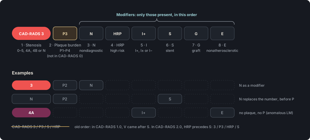
The CAD-RADS 2.0 code follows the order stenosis, plaque burden and modifiers, separated by slashes. After P come N, HRP, I, S, G and E, in that order, and only those that are present.
In practice. Write the stenosis category first, then P, which is omitted in CAD-RADS 0 or when there is no plaque. Then add the modifiers present in their fixed order. When N is the category it takes the place of the number, as in N/P2/S.
Pitfall. Writing /S before /HRP, as CAD-RADS 1.0 did with the former V modifier. In CAD-RADS 2.0, HRP is the second modifier, as in 3/P3/HRP/S.
Other sources. CAD-RADS 1.0 had four modifiers, in the order N, S, G and V, and did not include plaque burden. CAD-RADS 2.0 added P, I and E, and replaced V with HRP, which now precedes S. (CAD-RADS 1.0, Table 2, p. 271)
On the map. The map builds the code in the order P, N, HRP, I, S, G and E, and omits P, HRP and I in category 0. The impression in the template repeats the full code. Open the example on the map (CAD-RADS 3/P3/HRP/S) →
How the map's template words it
Left anterior descending (LAD): proximal segment: non-calcified plaque causing moderate stenosis (50-69%) with high-risk plaque features (low attenuation and positive remodeling); mid segment: partially calcified plaque resulting in mild stenosis (25-49%); the remainder is unremarkable. Branches D1, D2: no plaque.
Circumflex (LCx): proximal segment: calcified plaque causing minimal stenosis (1-24%); the rest of the vessel is unremarkable. 1st Obtuse marginal (OM1): calcified plaque causing minimal stenosis (1-24%); Branch OM2: no plaque.
Right coronary (RCA): proximal segment: patent stent, no significant restenosis; mid segment: partially calcified plaque producing mild stenosis (25-49%); the rest of the vessel is unremarkable. Branches AM, R-PDA, R-PLB: no plaque.
Source: CAD-RADS 2.0, Section 3.3, coding framework; SCCT 2026, «Impression» section, p. 36 · «First: modifier N (non-diagnostic) Second: modifier HRP (high-risk plaque) Third: modifier I (ischemia) Fourth: modifier S (stent) Fifth: modifier G (graft) Sixth: modifier E (exceptions)»
Figure 1. The code starts with the category, then plaque burden, and ends with the modifiers present, in a fixed order. When N is the category, it takes the place of the number.
Also seen as:CT-FFR, FFR-CT, FFRCT(Spelling used by HeartFlow; also written FFRct), CT-derived FFR
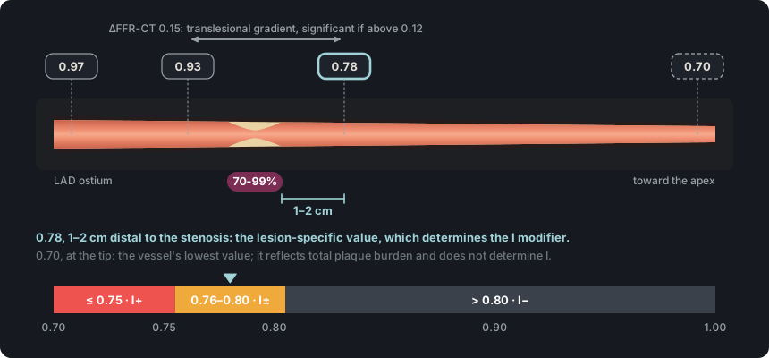
With CT-FFR, the I modifier is assigned from the lesion-specific value. A value of 0.75 or less is I+, 0.76 to 0.80 is I±, and above 0.80 is I−.
In practice. The value is read 1 to 2 cm distal to the stenosis, in a vessel large enough for intervention. With I±, the decision to proceed to invasive angiography depends on lesion location, symptoms and the translesional gradient, which is significant if it exceeds 0.12.
Pitfall. Assigning I from the lowest value in the vessel, which reflects total plaque burden. Use the value 1–2 cm distal to the lesion.
Other sources. For invasive FFR, the FAME trial used 0.80 or less as the threshold for stenting. With CT-FFR, CAD-RADS 2.0 reserves I+ for 0.75 or less and considers the 0.76 to 0.80 range borderline (I±). (Tonino 2009, FAME, Abstract)
On the map. Under «Modifiers P · I», each territory (LAD, LCx and RCA) accepts a CT-FFR value and classifies it as I+, I± or I−. The template records the value, for example «left anterior descending: CT-FFR 0.72». Open the example on the map (CAD-RADS 3/P1/I+) →
How the map's template words it
Left anterior descending (LAD): proximal segment: partially calcified plaque causing moderate stenosis (50-69%); the remainder is unremarkable. Branches D1, D2: no plaque.
Circumflex (LCx): distal segment: calcified plaque producing mild stenosis (25-49%); the rest of the vessel is unremarkable. Branches OM1, OM2: no plaque.
Source: CAD-RADS 2.0, Section 3.3.5.1 and Table 6 (Interpretation of CT-FFR) · «If there is presence of abnormal CT-FFR as defined as a lesion-specific value ≤ 0.75 in a vessel large enough for PCI the designation of “I+” (I=Ischemia) should be included.»
Figure 2. The I modifier is assigned from the value measured 1 to 2 cm distal to the stenosis. The lowest value, at the distal end of the vessel, reflects total plaque burden and should not drive referral to invasive angiography.
Also seen as:CT myocardial perfusion imaging, CT-MPI, myocardial CTP, stress CTP
On CT perfusion, a defect that is reversible or larger at stress than at rest is I+. No defect, or a fixed defect from infarction, is I−. A borderline result is I±.
In practice. Stress and rest are compared on short-axis images, using 5 to 8 mm averaged reconstructions and a narrow window, without MIP. A true defect persists across several phases of the cardiac cycle. If the defect is fixed, the prior infarct is documented in the impression.
Pitfall. Assigning I+ for myocardial hypoattenuation on resting CT angiography. The I modifier requires an ischemia test, either stress perfusion or CT-FFR.
On the map. With «CTP» selected, «Reversible» gives I+, and «Fixed» or «No defect» give I−. With a fixed defect, the impression in the template describes it as consistent with prior infarction and suggests viability assessment. Open the example on the map (CAD-RADS 4A/P1/I-) →
How the map's template words it
Right coronary (RCA): distal segment: partially calcified plaque causing severe stenosis (70-99%); the rest of the vessel is unremarkable. Branches AM, R-PDA, R-PLB: no plaque.
Impression Severe obstructive CAD. CAD-RADS 4A/P1/I-.
Source: CAD-RADS 2.0, Section 3.3.5.2 and Table 7 (Interpretation of Stress Myocardial CT Perfusion) · «If no ischemia is detected or if there is presence of a prior fixed myocardial infarct, the Modifier “I−” will be added to CAD-RADS.»
CAD-RADS 2.0 pairs each category with suggested management for the patient with stable chest pain. These are consensus suggestions that complement the impression and do not replace clinical judgment.
In practice. For categories 0 to 2 no further cardiac testing is suggested, and in 1 and 2 preventive therapy is adjusted to P. With 3, functional testing is considered; with 4A, invasive angiography or functional testing; and with 4B, invasive angiography is recommended.
Pitfall. Treating CAD-RADS 1 or 2 as settled without checking P. With P3 or P4, CAD-RADS 2.0 suggests intensive risk factor modification and preventive pharmacotherapy.
Other sources. In stable chest pain, the 2021 AHA/ACC guideline considers CT-FFR most useful for 40 to 90 % stenoses in proximal or mid segments. CAD-RADS 2.0 suggests functional assessment starting at CAD-RADS 3 (50–69 %). (AHA/ACC 2021 chest pain guideline, Section 5.1.3)
On the map. With the «Stable» clinical setting, the template adds the category's «Suggested management (CAD-RADS 2.0 consensus)», with layers for P, I and HRP, and a footer that leaves the decision to the treating physician. Open the example on the map (CAD-RADS 1/P3) →
How the map's template words it
Left main (LM): calcified plaque resulting in minimal stenosis (1-24%).
Left anterior descending (LAD): proximal segment: calcified plaque causing minimal stenosis (1-24%); mid segment: partially calcified plaque resulting in minimal stenosis (1-24%); the remainder is unremarkable. Branches D1, D2: no plaque.
Circumflex (LCx): proximal segment: calcified plaque causing minimal stenosis (1-24%); the rest of the vessel is unremarkable. Branches OM1, OM2: no plaque.
Right coronary (RCA): proximal segment: calcified plaque causing minimal stenosis (1-24%); the rest of the vessel is unremarkable. Branches AM, R-PDA, R-PLB: no plaque.
Source: CAD-RADS 2.0, Section 1 and Table 4; SCCT 2026, Table 6, p. 52 · «the clinical management suggestions provided by CAD-RADS should not replace clinical judgment, particularly as there are many patient-specific factors that may influence clinical management.»
Self-assessment
Short questions to check the key points. The answer appears when you choose.
1In a patient with stable chest pain, the left main has a 55 % stenosis, the mid LAD 40 % and the proximal RCA 30 %. There are no other lesions. Which CAD-RADS category is assigned?
Left main stenosis of 50 % or more is enough for CAD-RADS 4B, even if no lesion reaches 70 %. The LAD and RCA lesions are described in the findings. See the term
2The mid LAD has a moderate stenosis (50–69 %) and plaque burden is P2. Motion artifact prevents evaluation of the mid RCA. How is the study coded?
There is a stenosis of 50 % or more in an evaluable segment, so it is graded and N becomes a modifier after P. CAD-RADS N is used only when no evaluable segment reaches 50 %. See the term
3There is a patent stent in the proximal RCA. In the proximal LAD, a low-attenuation plaque with positive remodeling causes a moderate stenosis, and plaque burden is P3. What is the correct code?
P follows the category, and the modifiers follow in the order N, HRP, I, S, G and E. The legend of CAD-RADS 2.0 Figure 17 reverses S and HRP, but the ordered list takes precedence. See the term
4The mid LAD has a moderate stenosis. CT-FFR measured 1 to 2 cm distal to the lesion is 0.78, and the lowest value, at the apical end of the LAD, is 0.70. Which modifier is added?
The lesion-specific value, measured 1 to 2 cm distal to the stenosis, is used. A value from 0.76 to 0.80 is borderline. The lowest value in the vessel reflects total plaque burden. See the term
5The distal RCA has a severe stenosis (70–99 %). CT perfusion shows an inferior defect of the same size at stress and at rest, with wall thinning. How is ischemia coded?
A fixed defect from prior infarction, without inducible ischemia, is coded I−. CAD-RADS 2.0 reserves the I modifier for ischemia and asks that the infarct be documented in the impression. See the term
References
Rajiah PS, Alkadhi H, Andreini D, Bullock-Palmer RP, Chinnaiyan K, Chow B, et al. Interpretation and reporting of coronary computed tomographic angiography (2026 update): an expert consensus document of the Society of Cardiovascular Computed Tomography (SCCT). J Cardiovasc Comput Tomogr. 2026. In press. doi:10.1016/j.jcct.2026.08.014 Cited pages refer to the journal pre-proof PDF.
Cury RC, Leipsic J, Abbara S, Achenbach S, Berman D, Bittencourt M, et al. CAD-RADS 2.0 - 2022 Coronary Artery Disease-Reporting and Data System: an expert consensus document of the SCCT, ACC, ACR and NASCI. J Cardiovasc Comput Tomogr. 2022;16(6):536-57. doi:10.1016/j.jcct.2022.07.002
Koweek L, Achenbach S, Berman DS, Carr JJ, Cury RC, Ghoshhajra B, et al. Standardized medical terminology for cardiac computed tomography 2023 update: an expert consensus document of the SCCT, AAPM, ACR, NASCI and RSNA. J Cardiovasc Comput Tomogr. 2023;17(5):345-54. doi:10.1016/j.jcct.2023.06.002
Narula J, Chandrashekhar Y, Ahmadi A, Abbara S, Berman DS, Blankstein R, et al. SCCT 2021 expert consensus document on coronary computed tomographic angiography: a report of the Society of Cardiovascular Computed Tomography. J Cardiovasc Comput Tomogr. 2021;15(3):192-217. doi:10.1016/j.jcct.2020.11.001
Neves PO, Andrade J, Monção H. Coronary artery calcium score: current status. Radiol Bras. 2017;50(3):182-9.
Tamburino C, Tomasello SD, Capodanno D, Di Salvo ME, Marzà F, Galassi AR. Long-term follow-up after drug eluting stent implantation in left main trifurcations. EuroIntervention. 2009;5(4):432-7. doi:10.4244/eijv5i4a68
Kovacevic M, Burzotta F, Elharty S, Besis G, Aurigemma C, Romagnoli E, et al. Left main trifurcation and its percutaneous treatment: what is known so far? Circ Cardiovasc Interv. 2021;14(3):e009872. doi:10.1161/CIRCINTERVENTIONS.120.009872
Kim C, Park CH, Lee BY, et al. 2024 consensus statement on coronary stenosis and plaque evaluation in CT angiography from the Asian Society of Cardiovascular Imaging-Practical Tutorial (ASCI-PT). Korean J Radiol. 2024;25(4):331-42. doi:10.3348/kjr.2024.0112
Sources for the “Other sources” notes
Sianos G, Morel MA, Kappetein AP, et al. The SYNTAX Score: an angiographic tool grading the complexity of coronary artery disease. EuroIntervention. 2005;1(2):219-27.
Cury RC, Abbara S, Achenbach S, et al. CAD-RADS: Coronary Artery Disease – Reporting and Data System. J Cardiovasc Comput Tomogr. 2016;10(4):269-81. doi:10.1016/j.jcct.2016.04.005
Williams MC, Moss AJ, Dweck M, et al. Coronary artery plaque characteristics associated with adverse outcomes in the SCOT-HEART study. J Am Coll Cardiol. 2019;73(3):291-301. doi:10.1016/j.jacc.2018.10.066
Hecht HS, Blaha MJ, Kazerooni EA, et al. CAC-DRS: Coronary Artery Calcium Data and Reporting System. An expert consensus document of the Society of Cardiovascular Computed Tomography (SCCT). J Cardiovasc Comput Tomogr. 2018;12(3):185-91. doi:10.1016/j.jcct.2018.03.008
Lawton JS, Tamis-Holland JE, Bangalore S, et al. 2021 ACC/AHA/SCAI guideline for coronary artery revascularization: a report of the American College of Cardiology/American Heart Association Joint Committee on Clinical Practice Guidelines. J Am Coll Cardiol. 2022;79(2):e21-e129. doi:10.1016/j.jacc.2021.09.006
Galassi AR, Werner GS, Boukhris M, et al. Percutaneous recanalisation of chronic total occlusions: 2019 consensus document from the EuroCTO Club. EuroIntervention. 2019;15(2):198-208. doi:10.4244/EIJ-D-18-00826
Tonino PA, De Bruyne B, Pijls NH, et al. Fractional flow reserve versus angiography for guiding percutaneous coronary intervention. N Engl J Med. 2009;360(3):213-24. doi:10.1056/NEJMoa0807611
Gulati M, Levy PD, Mukherjee D, et al. 2021 AHA/ACC/ASE/CHEST/SAEM/SCCT/SCMR Guideline for the Evaluation and Diagnosis of Chest Pain: A Report of the American College of Cardiology/American Heart Association Joint Committee on Clinical Practice Guidelines. Circulation. 2021;144(22):e368-e454. doi:10.1161/CIR.0000000000001029