Nonatherosclerotic coronary findings and modifier E
David F. Cuevas Cueto, MD · Updated September 27, 2026 · Sources: SCCT 2026 and CAD-RADS 2.0
This section covers the most common nonatherosclerotic coronary findings on CCTA, such as myocardial bridging, anomalous origin and course, and aneurysm or ectasia. CAD-RADS 2.0 flags the nonatherosclerotic findings on its list with modifier E, which is added to the stenosis category without replacing it.
Key points
A myocardial bridge is described by its location, depth and length. It is deep when 2 mm or more of myocardium covers the artery, and long when the tunnel measures 25 mm or more.
When only one phase is acquired, systolic compression of the bridge cannot be assessed reliably, and that limitation should be kept in mind when reporting it.
Neither CAD-RADS 2.0 nor SCCT 2026 names myocardial bridging in the modifier E list, which is not exhaustive, so adding E for a bridge is a matter of interpretation.
An anomalous coronary artery is described by its origin, course, luminal caliber and termination; the interarterial course is the high-risk one, especially with an intramural segment.
An aneurysm is a dilation to more than 1.5 times the reference diameter that involves less than half of the vessel; when the dilation involves more than half, it is called ectasia.
Modifier E goes at the end of the code, after N, HRP, I, S and G, and when no segment has plaque the code has no P.
Table 1. Courses of an anomalous coronary artery. SCCT 2026 describes four; the retrocardiac course comes from publications on single coronary artery.
Course
Path
Risk reported in the sources
On the map
Interarterial
Between the aortic root and the pulmonary trunk
High, especially with an intramural segment
«Interarterial»
Transseptal (subpulmonic)
Beneath the pulmonary valve, within the septum
Lower than the interarterial course
Not available
Prepulmonic
Anterior to the pulmonary trunk
Generally benign
«Prepulmonary»
Retroaortic
Posterior to the aortic root
Generally benign
«Retroaortic»
Retrocardiac
Posterior to the heart
Generally benign
Not available
Source: SCCT 2026, pp. 20–21 (courses, high risk of the interarterial course and less compression of the transseptal course); Cheezum 2017, J Am Coll Cardiol, abstract (most subtypes are benign and sudden death is associated with the interarterial course); Sheng 2021 (retrocardiac course, generally benign).
Also seen as:myocardial bridge, MB, tunneled coronary artery, mural coronary artery(Mostly pathology literature)
A segment of an epicardial artery runs through a tunnel within the myocardium, and the muscle band covering it is the bridge. The LAD is the most commonly affected artery, particularly its mid segment.
In practice. It is identified on the curved reconstruction of the LAD and confirmed on cross-sectional views, where the tunneled segment is surrounded by myocardium. In the findings, the vessel and segment are named, and depth, length and systolic compression are described.
Pitfall. Reviewing only the tunnel and skipping the segment proximal to it. Plaque forms mainly just proximal to the bridge, and the tunneled segment is usually spared.
Other sources. SCCT 2014 listed myocardial bridging among the potentially relevant anatomical variants, together with absent left main. Because of its high prevalence, some authors consider it a variant rather than a true anomaly, although most classifications count it among anomalies of course. (SCCT 2014, section 10.6.2, p. 352; Genoud 2026, section 4.2 and Table 2)
On the map. A bridge can be added only to the mid LAD, from the «Myocardial bridge» section of its editor, with «Add bridge». Plaque cannot be placed in the tunnel, and the bridge does not change the category. The app adds E by convention. Open the example on the map (CAD-RADS 3/P1/E) →
How the map's template words it
Left anterior descending (LAD): proximal segment: non-calcified plaque causing moderate stenosis (50-69%); the remainder is unremarkable. Myocardial bridge of the mid segment, with an intramyocardial course (superficial) and ~20% systolic compression. Branches D1, D2: no plaque.
Source: SCCT 2026, text, nonatherosclerotic coronary artery abnormalities (section j), p. 21 · «Myocardial bridging is characterized by encasement of coronary arteries by myocardial fibers, with a tunneled intramyocardial segment and a myocardial bridge that overlies the artery.»
Myocardial bridge depth, length and systolic compression#
Also seen as:milking effect(Invasive angiography term), deep myocardial bridge, superficial myocardial bridge, myocardial bridge thickness
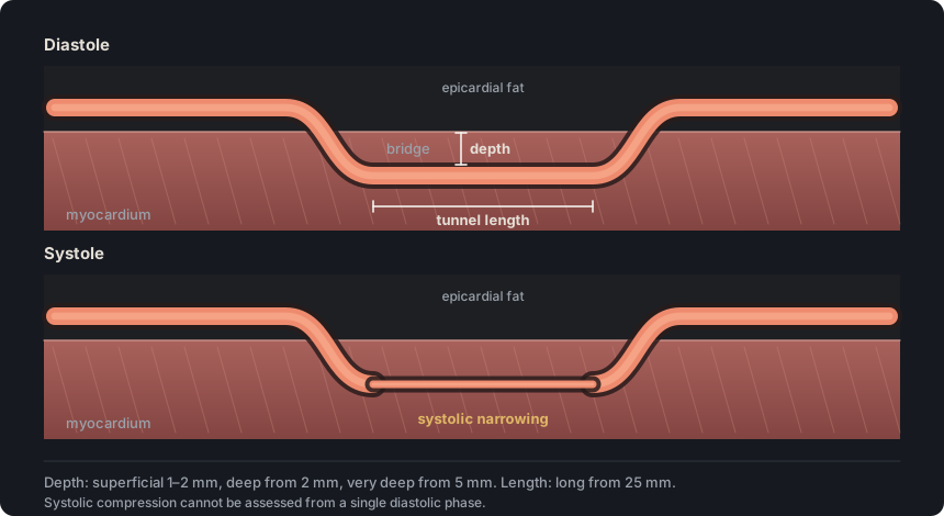
SCCT 2026 classifies the bridge by depth and length. It is superficial with 1 to 2 mm of overlying myocardium, deep with 2 mm or more, very deep with 5 mm or more, and long when it measures 25 mm or more.
In practice. The thickness of myocardium over the artery is measured on the cross-sectional view, and the tunnel length on the curved reconstruction. Systolic compression, assessed by comparing systole with diastole, may be absent, less than 50 %, or 50 % or more.
Pitfall. Reporting a systolic compression percentage when only a diastolic phase was acquired. SCCT 2026 cautions that anatomical assessment is limited with a single phase.
Other sources. On invasive angiography, the Noble classification grades systolic compression of the LAD in three levels. Grade 1 is less than 50 %, grade 2 is 50 to 75 % and grade 3 exceeds 75 %. On CCTA, SCCT 2026 divides systolic compression into absent, less than 50 %, or 50 % or more. (Noble 1976, cited by Xu 2022, methods; SCCT 2026, p. 21)
On the map. «Deep» and «Superficial» have no millimeter threshold, and «Tunnel length» is set as a fraction of the segment and is not printed. The template always includes the compression percentage, except in a nondiagnostic (N) segment. Open the example on the map (CAD-RADS 0/E) →
How the map's template words it
Left anterior descending (LAD): no atherosclerotic disease. Myocardial bridge of the mid segment, with an intramyocardial course (deep) and ~55% systolic compression. Branches D1, D2: no plaque.
Impression No coronary atherosclerosis. CAD-RADS 0/E.
Source: SCCT 2026, text, nonatherosclerotic coronary artery abnormalities (section j), p. 21 · «superficial (1-2 mm), deep (≥ 2 mm), very deep (≥ 5 mm) and short (<25 mm) or long (≥ 25 mm)»
Figure 1. The bridge is measured by the thickness of myocardium covering the artery and by the length of the tunnel. Compression becomes apparent when systole is compared with diastole, and with a single phase its assessment is limited.
Anomalous origin of a coronary artery AAOCA · ALCAPA · ARCAPA#
Also seen as:ACAOS(Origin from the opposite sinus of Valsalva), ACAPA(Origin from the pulmonary artery), anomalous aortic origin of a coronary artery, Bland-White-Garland syndrome(Eponym for ALCAPA), anomalous coronary artery origin
A coronary artery has an anomalous origin when it arises outside its usual site. It may arise from the opposite or the noncoronary sinus of Valsalva, in a high, low or commissural position, or from the pulmonary artery.
In practice. Each artery is followed from its ostium on axial images and oblique reconstructions of the aortic root. SCCT 2026 recommends describing origin, morphology, course, luminal caliber and termination, and the relationship to adjacent structures.
Pitfall. Reporting a conus branch that arises directly from the right sinus as anomalous. SCCT 2026 lists that origin as one of the two usual origins of the conus branch.
Other sources. SCCT 2021 uses ACAOS for origin from the opposite sinus of Valsalva. The ESC 2020 guideline uses AAOCA for anomalous aortic origin and ACAPA for origin from the pulmonary artery, with its ALCAPA and ARCAPA forms. (SCCT 2021, section 4.10, p. 209; ESC 2020, section 4.17.1)
On the map. In the «One vessel» tab of «Anomalous origin (E)», the user selects the vessel (RCA, LM, LAD or LCx) and its origin: «R sinus», «L sinus» or «PA». There is no noncoronary sinus option. Open the example on the map (CAD-RADS 0/E) →
How the map's template words it
Origin and course: Circumflex (LCx) with an anomalous origin from the right coronary sinus, a retroaortic course. Dominance: right.
Impression No coronary atherosclerosis. CAD-RADS 0/E.
Source: SCCT 2026, text, nonatherosclerotic coronary artery abnormalities (section j); conus branch in coronary anatomy, p. 12 · «Anomalous origin could be from the opposite or non-coronary sinus of Valsalva or from pulmonary arteries.»
Also seen as:intraseptal course(Other name for the transseptal course), precardiac course(Other name for the prepulmonic course), anomalous coronary course
The course indicates where the anomalous artery crosses the base of the heart. SCCT 2026 recognizes four courses: interarterial, retroaortic, transseptal or subpulmonic, and prepulmonic.
In practice. The proximal segment is followed on axial and oblique images until it reaches its groove. The interarterial course passes between the aortic root and the pulmonary trunk and is high risk. The transseptal course is compressed less because it lies between the aorta and the contracted right ventricular outflow tract.
Pitfall. Confusing the transseptal course with the interarterial course. The transseptal course passes beneath the pulmonary valve, within the septum, and not between the aorta and the pulmonary trunk.
Other sources. Some reviews describe five courses, adding the retrocardiac course, which passes behind the heart. Other names found in the literature are intraseptal for transseptal and precardiac for prepulmonic. This glossary follows the four courses of SCCT 2026. (Genoud 2026, section 4.1.3; Evangelista 2024; Karatoprak 2026, methods)
On the map. In «One vessel», the user selects «Interarterial», «Prepulmonary» or «Retroaortic», and the app suggests one based on the vessel and sinus. There is no transseptal option. The template describes the interarterial course as a high-risk variant. Open the example on the map (CAD-RADS 0/E) →
How the map's template words it
Origin and course: Right coronary (RCA) with an anomalous origin from the left coronary sinus, an interarterial course. Dominance: right.
Impression No coronary atherosclerosis. CAD-RADS 0/E.
Source: SCCT 2026, text, nonatherosclerotic coronary artery abnormalities (section j), pp. 20–21 · «Anomalous courses could be interarterial, retroaortic, transseptal (subpulmonic) or prepulmonic»
The intramural segment is the proximal portion of the anomalous artery that runs within the aortic wall before separating from it. It increases the risk associated with an interarterial course.
In practice. CT does not show it directly. It is inferred from an acute take-off angle, segmental narrowing with a slit-like orifice on the cross-sectional view, and incomplete encasement by pericoronary fat on the side facing the aortic lumen. When recognized, its length is measured.
Pitfall. Assessing the shape of the proximal lumen on the axial plane, which cuts the vessel obliquely. The slit-like orifice is seen on the cross-sectional view.
Other sources. In the Angelini classification, «intramural coronary artery» is another name for myocardial bridging. In this glossary, as in SCCT 2026, the term intramural is reserved for the portion of the anomalous artery that runs within the aortic wall, and the tunneled segment of a bridge is called intramyocardial. (Ganga 2021, Table 1; Evangelista 2024; Genoud 2026, Figure 5)
On the map. In «High-risk features», the user selects «Intramural course» and enters the length in «Intramural length (mm)». The template prints it as «intramural segment (~6 mm)». The option is available only with an aortic origin. Open the example on the map (CAD-RADS 0/E) →
How the map's template words it
Origin and course: Right coronary (RCA) with an anomalous origin from the left coronary sinus, an interarterial course. Dominance: right.
Impression No coronary atherosclerosis. CAD-RADS 0/E.
Source: SCCT 2026, text, nonatherosclerotic coronary artery abnormalities (section j), p. 20 · «An intramural segment is not directly visualized, but can be inferred by an acute takeoff angle, segmental narrowing with a slit-like orifice at cross-section»
High-risk anatomy of an anomalous coronary artery#
Also seen as:high-risk features of AAOCA
An interarterial course, an intramural segment, a slit-like orifice, an acute take-off angle, proximal narrowing and a high origin are considered high risk because they are associated with ischemia.
In practice. Once the course is defined, the ostium and proximal segment are reviewed on cross-sectional views. The shape of the orifice, the take-off angle, the length of the narrowing and the height of the ostium relative to the sinotubular junction are reported.
Pitfall. Giving the same weight to an RCA from the left sinus as to a left coronary artery from the right sinus. ESC 2020 considers the latter more malignant.
Other sources. The ESC 2020 guideline considers an ostium located more than 1 cm above the sinotubular junction high risk. Other authors call any ostium above that junction a high origin, and SCCT 2026 does not set a distance. (ESC 2020, section 4.17.2 and note c; Karatoprak 2026, methods)
On the map. With an aortic origin, «High-risk features» offers «Slit-like ostium / acute take-off», «Intramural course» and «High ostial origin». The template lists them in the impression, and none changes the category. Open the example on the map (CAD-RADS 0/E) →
How the map's template words it
Origin and course: Left main (LM) with an anomalous origin from the right coronary sinus, an interarterial course. Dominance: right.
Impression No coronary atherosclerosis. CAD-RADS 0/E.
Source: SCCT 2026, text, nonatherosclerotic coronary artery abnormalities (section j), p. 20 · «An interarterial course between the aortic root and pulmonary artery is considered a high-risk anatomy, particularly when combined with an intramural course.»
Also seen as:CAA, CAE, coronary aneurysm, coronary ectasia, aneurysmal coronary artery disease(Older term)
A coronary aneurysm is a segment with a diameter more than 1.5 times that of the adjacent normal segment, involving less than 50 % of the vessel length. If it involves more, it is ectasia.
In practice. The maximum diameter is measured on the cross-sectional view and compared with the adjacent normal segment or with the largest coronary artery. In a saccular aneurysm the transverse diameter is greater than the longitudinal diameter; in a fusiform aneurysm the longitudinal diameter is greater.
Pitfall. Calling a dilation that involves more than half of the vessel an aneurysm. That diffuse dilation is ectasia, which SCCT 2026 attributes mainly to atherosclerosis.
On the map. The map has no control for aneurysm or ectasia, so the app does not add E even though aneurysm is on the CAD-RADS 2.0 list. Both are typed by hand into the template.
Source: SCCT 2026, text, nonatherosclerotic coronary artery abnormalities (section j), p. 22 · «Coronary artery aneurysm is defined as a coronary artery segment with a diameter > 1.5 times the diameter of normal adjacent coronary artery segments or the largest coronary artery»
Modifier E flags a nonatherosclerotic coronary finding. It is written at the end of the CAD-RADS code, after N, HRP, I, S and G, and accompanies the category without replacing it.
In practice. Table 3 of CAD-RADS 2.0 is not an exhaustive list. It includes dissection, anomalous origin, aneurysm or pseudoaneurysm, vasculitis, fistula, extrinsic compression, arteriovenous malformation and other causes. Nonatherosclerotic narrowing is graded, and an interarterial anomaly with moderate stenosis is CAD-RADS 3/E.
Pitfall. Adding P to the code of an anomaly when no segment has plaque. CAD-RADS 2.0 omits the P in its example of an interarterial anomaly without plaque.
Other sources. CAD-RADS 1.0 had only modifiers N, S, G and V, so a nonatherosclerotic finding had no code of its own. CAD-RADS 2.0 introduced modifiers I and E and renamed modifier V (vulnerable plaque) as HRP (high-risk plaque). (CAD-RADS 1.0, Table 1, p. 271; CAD-RADS 2.0, sections 3.3 and 3.3.6)
On the map. The app adds E for an anomaly marked in «One vessel», a myocardial bridge or a single coronary artery. It does not grade narrowing without plaque, and for other exceptions the E is typed by hand. Open the example on the map (CAD-RADS 2/P1/HRP/E) →
How the map's template words it
Origin and course: Circumflex (LCx) with an anomalous origin from the right coronary sinus, a retroaortic course. Dominance: right.
Left anterior descending (LAD): proximal segment: non-calcified plaque causing mild stenosis (25-49%) with high-risk plaque features (low attenuation and positive remodeling); the remainder is unremarkable. Branches D1, D2: no plaque.
Source: CAD-RADS 2.0, section 3.3.6 (Modifier E = exceptions) and Table 3 · «a modifier “E” is used to account for any non-atherosclerotic narrowing of the coronary arteries and should be added at the end of the score as a modifier»
Self-assessment
Short questions to check the key points. The answer appears when you choose.
1The RCA arises from the left coronary sinus with an interarterial course. In its proximal segment, compression between the aorta and the pulmonary trunk causes a moderate stenosis. No segment has plaque. How is this coded?
CAD-RADS 2.0 gives an interarterial anomaly with moderate stenosis as an example and codes it 3/E. Because no segment has plaque, the code has no P. See the term
2In the mid LAD, a 30 mm segment runs within the myocardium, covered by a 3 mm muscle layer on the cross-sectional view. How is the bridge classified according to SCCT 2026?
A bridge is deep with 2 mm or more of overlying myocardium and very deep with 5 mm or more, so 3 mm makes it deep. It measures 30 mm, and a bridge of 25 mm or more is classified as long. See the term
3The LAD arises from the right coronary sinus. Its proximal segment passes beneath the pulmonary valve, within the interventricular septum, and resurfaces in the anterior interventricular groove. How is the course described?
The transseptal or subpulmonic course runs within the septum, beneath the pulmonary valve. SCCT 2026 notes that it is compressed less than the interarterial course, which passes between the aorta and the pulmonary trunk. See the term
4An anomalous RCA arises from the left sinus with an interarterial course. On the cross-sectional view of its proximal portion, the lumen is an oval slit, the take-off forms an acute angle, and pericoronary fat is missing on the aortic side. What do these signs suggest?
SCCT 2026 explains that an intramural segment is not directly visualized. It is inferred from an acute take-off angle, a slit-like orifice and incomplete encasement by pericoronary fat on the side facing the aortic lumen. See the term
5The RCA measures 7 mm along 80 % of its length. Its distal segment, of normal caliber, measures 4 mm. How is the dilation classified?
The diameter is 1.75 times the reference diameter, which exceeds 1.5. Because the dilation involves more than 50 % of the vessel, SCCT 2026 calls it ectasia rather than aneurysm. See the term
References
Rajiah PS, Alkadhi H, Andreini D, Bullock-Palmer RP, Chinnaiyan K, Chow B, et al. Interpretation and reporting of coronary computed tomographic angiography (2026 update): an expert consensus document of the Society of Cardiovascular Computed Tomography (SCCT). J Cardiovasc Comput Tomogr. 2026. In press. doi:10.1016/j.jcct.2026.08.014 Cited pages refer to the journal pre-proof PDF.
Cury RC, Leipsic J, Abbara S, Achenbach S, Berman D, Bittencourt M, et al. CAD-RADS 2.0 - 2022 Coronary Artery Disease-Reporting and Data System: an expert consensus document of the SCCT, ACC, ACR and NASCI. J Cardiovasc Comput Tomogr. 2022;16(6):536-57. doi:10.1016/j.jcct.2022.07.002
Koweek L, Achenbach S, Berman DS, Carr JJ, Cury RC, Ghoshhajra B, et al. Standardized medical terminology for cardiac computed tomography 2023 update: an expert consensus document of the SCCT, AAPM, ACR, NASCI and RSNA. J Cardiovasc Comput Tomogr. 2023;17(5):345-54. doi:10.1016/j.jcct.2023.06.002
Narula J, Chandrashekhar Y, Ahmadi A, Abbara S, Berman DS, Blankstein R, et al. SCCT 2021 expert consensus document on coronary computed tomographic angiography: a report of the Society of Cardiovascular Computed Tomography. J Cardiovasc Comput Tomogr. 2021;15(3):192-217. doi:10.1016/j.jcct.2020.11.001
Neves PO, Andrade J, Monção H. Coronary artery calcium score: current status. Radiol Bras. 2017;50(3):182-9.
Tamburino C, Tomasello SD, Capodanno D, Di Salvo ME, Marzà F, Galassi AR. Long-term follow-up after drug eluting stent implantation in left main trifurcations. EuroIntervention. 2009;5(4):432-7. doi:10.4244/eijv5i4a68
Kovacevic M, Burzotta F, Elharty S, Besis G, Aurigemma C, Romagnoli E, et al. Left main trifurcation and its percutaneous treatment: what is known so far? Circ Cardiovasc Interv. 2021;14(3):e009872. doi:10.1161/CIRCINTERVENTIONS.120.009872
Kim C, Park CH, Lee BY, et al. 2024 consensus statement on coronary stenosis and plaque evaluation in CT angiography from the Asian Society of Cardiovascular Imaging-Practical Tutorial (ASCI-PT). Korean J Radiol. 2024;25(4):331-42. doi:10.3348/kjr.2024.0112
Sources for the “Other sources” notes
Leipsic J, Abbara S, Achenbach S, Cury R, Earls JP, Mancini GBJ, et al. SCCT guidelines for the interpretation and reporting of coronary CT angiography: a report of the Society of Cardiovascular Computed Tomography Guidelines Committee. J Cardiovasc Comput Tomogr. 2014;8(5):342-58. doi:10.1016/j.jcct.2014.07.003
Cury RC, Abbara S, Achenbach S, et al. CAD-RADS: Coronary Artery Disease – Reporting and Data System. J Cardiovasc Comput Tomogr. 2016;10(4):269-81. doi:10.1016/j.jcct.2016.04.005
Noble J, Bourassa MG, Petitclerc R, et al. Myocardial bridging and milking effect of the left anterior descending coronary artery: normal variant or obstruction? Am J Cardiol. 1976;37(7):993-9. doi:10.1016/0002-9149(76)90414-8
Xu T, You W, Wu Z, et al. Retrospective analysis of OCT on MB characteristics and 1-year follow-up of the ISR incidence after the DES implantation in patients with MB. Sci Rep. 2022;12(1):534. doi:10.1038/s41598-021-04579-9
Narula J, Chandrashekhar Y, Ahmadi A, et al. SCCT 2021 expert consensus document on coronary computed tomographic angiography: a report of the Society of Cardiovascular Computed Tomography. J Cardiovasc Comput Tomogr. 2021;15(3):192-217. doi:10.1016/j.jcct.2020.11.001
Baumgartner H, De Backer J, Babu-Narayan SV, et al. 2020 ESC Guidelines for the management of adult congenital heart disease. Eur Heart J. 2021;42(6):563-645. doi:10.1093/eurheartj/ehaa554
Genoud A, Portugal I, Murith N, et al. Coronary artery anomalies revisited: description of the types, pathophysiology and treatment options based on latest guidelines. J Clin Med. 2026;15(13):4959. doi:10.3390/jcm15134959
Evangelista M, Ferrero P, D'Aiello AF, et al. Coronary artery anomalies: what are they? when to suspect? how to treat?—a narrative review. Transl Pediatr. 2024;13(7):1242-57. doi:10.21037/tp-24-30
Karatoprak NB, Karatoprak S, Şahin Ö. Coronary artery origin anomalies on coronary CT angiography: a single-center tertiary-care cohort. J Clin Pract Res. 2026;48(2):137-44. doi:10.14744/cpr.2026.27967
Ganga KP, Goyal A, Ojha V, et al. Prevalence rates of congenital coronary anomalies and coronary variations in adult Indian population using dual-source computed tomography coronary angiography: analysis of regional distribution of coronary anomalies and the need for standardized reporting formats. Indian J Radiol Imaging. 2021;31(1):138-49. doi:10.1055/s-0041-1730135