Coronary stenosis: grading, reference diameter and total occlusion
David F. Cuevas Cueto, MD · Updated September 27, 2026 · Sources: SCCT 2026 and CAD-RADS 2.0
Stenosis is graded by the reduction in luminal diameter relative to a normal reference segment and is reported as a term with its range. The patient's CAD-RADS category is based on the most severe stenosis, and a left main stenosis of 50 % or more, or three-vessel disease, raises it to 4B. Definitions follow the SCCT 2026 consensus and CAD-RADS 2.0.
Key points
Stenosis is graded by diameter, comparing the minimal lumen with the lumen of a nearby normal segment, never with the outer contour of a remodeled plaque.
The grades are minimal (1–24 %), mild (25–49 %), moderate (50–69 %), severe (70–99 %) and total occlusion (100 %), and the report always gives the term with its range.
A visual estimate of the stenosis range is sufficient for the clinical report; quantitative measurement is not a routine requirement and varies by about ±25 % compared with invasive angiography.
A segment with no luminal opacification is a total occlusion even when there is distal contrast, and chronicity is not assumed but inferred from collaterals, calcium or length.
A stenosis is obstructive at 50 % or more and severe at 70 % or more, but in the left main a stenosis of 50 % or more is already CAD-RADS 4B.
Each stenosis is described by segment, with its anatomical name, plaque type and grade; in serial or diffuse disease, the most severe stenosis sets the category.
Table 1. Coronary stenosis grading scale by diameter. The category shown applies to the patient when that grade is the most severe stenosis; a left main stenosis of 50 % or more is CAD-RADS 4B.
Grade
Term
Range
Meaning
0
No visible stenosis
0 %
No luminal narrowing. Without plaque, CAD-RADS 0; with plaque, CAD-RADS 1
1
Minimal
1–24 %
Nonobstructive (CAD-RADS 1)
2
Mild
25–49 %
Nonobstructive (CAD-RADS 2)
3
Moderate
50–69 %
Obstructive (CAD-RADS 3; 4B if in the left main)
4
Severe
70–99 %
Obstructive (CAD-RADS 4A; 4B if in the left main or in three vessels)
5
Total occlusion
100 %
Nonopacified segment, acute or chronic (CAD-RADS 5)
Source: SCCT 2026, text p. 15 and Table 5, p. 51; CAD-RADS 2.0, Tables 1 and 4. Table 5 gives «minimal (1–25 %)»; the SCCT 2026 text and CAD-RADS 2.0 give «1–24 %».
Also seen as:percent diameter stenosis, DS, luminal diameter reduction
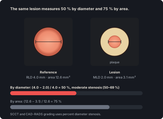
Diameter stenosis is the percent reduction in luminal diameter at the narrowest point relative to a normal reference segment. SCCT and CAD-RADS grade stenosis by diameter, not by area.
In practice. Maximal stenosis is estimated in transverse and longitudinal views, because curved MPR can underestimate an eccentric stenosis. If measured, it is calculated as (RLD − MLD) / RLD × 100, where RLD is the reference luminal diameter and MLD the minimal luminal diameter.
Pitfall. Reporting percent area stenosis as if it were diameter stenosis. Area gives higher figures for the same lesion, and the CAD-RADS scale applies to diameter.
Other sources. SCCT 2014 treated area stenosis as optional additional information. In a circular lumen, 50 % diameter stenosis equals 75 % area stenosis, so the CAD-RADS ranges do not apply to that figure. (SCCT 2014, section 5.4, p. 346; SCCT 2024 quantification, Table 2, p. 433; CAD-RADS 2.0, Table 1)
On the map. The 0–5 buttons and the «Stenosis grade» slider record the percentage on the CAD-RADS diameter scale, and each segment takes its highest-grade lesion. There is no field for area stenosis.
Source: SCCT 2026, qualitative stenosis grading, p. 15; curved MPR, p. 9; formula under quantitative grading, p. 16 · «A qualitative interpretation of the extent of coronary artery stenosis should be made in transverse and longitudinal views as an estimated maximal percent diameter stenosis.»
Figure 1. The same lesion measures 50 % by diameter and 75 % by area, because area is proportional to the square of the diameter. The SCCT and CAD-RADS scale applies to percent diameter stenosis.
The reference luminal diameter is measured in the lumen of a normal segment near the stenosis, proximal or distal, or taken as the average of both. It is the denominator of percent stenosis.
In practice. Choose the most normal cross-section within 10 mm of the lesion, with no branch vessels in between. For an ostial lesion, use the nearest distal reference. In the left main and in diffuse disease, normal reference diameters can be used.
Pitfall. Comparing the lumen with the outer contour of a positively remodeled plaque overestimates stenosis. The minimal lumen is compared with the lumen of a normal segment.
Other sources. The 2024 ASCI-PT consensus prefers the most normal-appearing site immediately proximal to the plaque. If the proximal segment is unsuitable, as in an ostial lesion, it uses the nearest distal reference. An interpolated reference remains an option for comparison with quantitative coronary angiography. (Kim 2024, ASCI-PT, topic 7 and Table 4)
On the map. The map does not measure diameters. The percentage recorded with the buttons or the slider is the one the radiologist estimated or measured at the workstation.
Source: SCCT 2026, qualitative (p. 15) and quantitative (pp. 16–17) stenosis grading; overestimation, p. 29 · «Qualitatively, the luminal diameter at a stenosis should be compared with the luminal diameter of a normal reference vessel segment distal and/or proximal to the stenosis.»
SCCT 2026 and CAD-RADS 2.0 divide diameter stenosis into six grades: 0 %, minimal (1–24 %), mild (25–49 %), moderate (50–69 %), severe (70–99 %) and total occlusion (100 %).
In practice. In the report, the term is written with its range, for example «moderate stenosis (50–69 %)», so that it is compatible with invasive angiography categories. The CAD-RADS category is based on the patient's most severe stenosis.
Pitfall. Writing only «moderate stenosis» without the range, or using words outside the scale, such as «critical». Write a term from the scale followed by its range.
Other sources. SCCT 2009 allowed an optional scale in which mild was less than 39 % and moderate 40 to 69 %. Some studies also use other cutoffs for mild and minimal. Before comparing an article with this scale, check which cutoffs it used. (Raff 2009, SCCT, optional quantitative scale; Kim 2024, ASCI-PT, topic 6 and Table 3)
On the map. The editor's 0–5 buttons store a representative value for each range (0, 15, 38, 60, 85 or 100 %), and the template writes the term with its range, for example «moderate stenosis (50-69%)». Open the example on the map (CAD-RADS 4A/P2) →
How the map's template words it
Left anterior descending (LAD): proximal segment: calcified plaque causing minimal stenosis (1-24%); mid segment: partially calcified plaque resulting in mild stenosis (25-49%); the remainder is unremarkable. Branches D1, D2: no plaque.
Circumflex (LCx): proximal segment: non-calcified plaque causing moderate stenosis (50-69%); the rest of the vessel is unremarkable. Branches OM1, OM2: no plaque.
Right coronary (RCA): mid segment: partially calcified plaque producing severe stenosis (70-99%); the rest of the vessel is unremarkable. Branches AM, R-PDA, R-PLB: no plaque.
Impression Severe obstructive CAD. CAD-RADS 4A/P2.
Source: SCCT 2026, qualitative stenosis grading, p. 15 · «This stenosis can be categorized as minimal (1-24 %), mild (25-49 %), moderate (50-69 %), severe (70-99 %) and total occlusion (100 %)»
Also seen as:complete occlusion, 100% stenosis, occluded segment
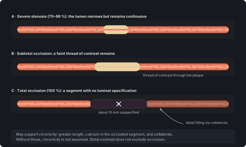
A total occlusion is a coronary segment with no luminal opacification, equivalent to 100 % stenosis. A single total occlusion, acute or chronic, makes the study CAD-RADS 5.
In practice. Follow the segment on curved MPR and transverse views. If a thread of contrast crosses the lesion, it is a subtotal occlusion. Distal contrast does not exclude occlusion, because the delayed acquisition allows time for collateral filling.
Pitfall. Writing «chronic occlusion» or «CTO» only because the segment is occluded. Chronicity is inferred separately, from findings such as collaterals, calcium or greater length.
Other sources. In the catheterization laboratory, chronic total occlusion requires absent antegrade flow through the lesion and a probable or documented duration of three months or more. On CCTA, length, calcium and collaterals only suggest chronicity. (Galassi 2024, EAPCI consensus on chronic total occlusions, introduction; SCCT 2026, p. 18; SCCT 2024 quantification, Table 4, p. 437)
On the map. Button 5 sets the lesion to 100 %, the template writes «total occlusion (100%)» and the category is CAD-RADS 5. The template adds «consistent with chronic total occlusion» only when a collateral reaching the occluded territory is turned on under «Collateral circulation». Open the example on the map (CAD-RADS 5/P1) →
How the map's template words it
Right coronary (RCA): mid segment: non-calcified plaque, total occlusion (100%); the rest of the vessel is unremarkable. Branches AM, R-PDA, R-PLB: no plaque.
Distal reconstitution of the occluded RCA via collaterals (from the LAD · apical PDA–distal LAD anastomosis), consistent with chronic total occlusion.
Impression Total occlusion (100%) of at least one vessel. CAD-RADS 5/P1.
Source: SCCT 2026, coronary occlusion (total, subtotal and features of chronicity), p. 18 · «Total coronary occlusion is defined by complete nonopacification of a coronary artery segment»
Figure 2. Total occlusion is defined by absent opacification of a segment, and distal contrast does not exclude it because it can arrive through collaterals. A thread of contrast through the lesion indicates subtotal occlusion.
Obstructive and nonobstructive stenosis ≥50 % · ≥70 %#
Also seen as:obstructive CAD, nonobstructive CAD, non-obstructive coronary artery disease
A stenosis of 50 % or more is called obstructive, and one below 50 % nonobstructive. The 70 % cutoff marks severe stenosis and defines obstructive three-vessel disease.
In practice. The 50 % cutoff separates CAD-RADS 2 from CAD-RADS 3, for which CAD-RADS 2.0 suggests considering functional testing. The 70 % cutoff separates CAD-RADS 3 from 4A. A left main stenosis of 50 % or more, or three-vessel disease with 70 % or more in each vessel, is CAD-RADS 4B.
Pitfall. Classifying a 50 to 69 % left main stenosis as CAD-RADS 3. In the left main, a stenosis of 50 % or more is CAD-RADS 4B.
Other sources. The 2021 ACC/AHA/SCAI revascularization guideline defines a significant angiographic stenosis as 70 % or more, or 50 % or more in the left main, and an intermediate stenosis as 40–69 %. The glossary follows the 50 % cutoff of CAD-RADS 2.0. (Lawton 2022, ACC/AHA/SCAI revascularization guideline, section 4.1)
On the map. The impression calls CAD-RADS 1 and 2 «nonobstructive» and lists the vessels with obstructive disease (≥50 %). A left main stenosis of 50 % or more gives CAD-RADS 4B even without a severe stenosis. Open the example on the map (CAD-RADS 3/P2) →
How the map's template words it
Left anterior descending (LAD): proximal segment: partially calcified plaque causing moderate stenosis (50-69%); the remainder is unremarkable. Branches D1, D2: no plaque.
Circumflex (LCx): proximal segment: partially calcified plaque causing moderate stenosis (50-69%); the rest of the vessel is unremarkable. Branches OM1, OM2: no plaque.
Right coronary (RCA): mid segment: partially calcified plaque producing moderate stenosis (50-69%); the rest of the vessel is unremarkable. Branches AM, R-PDA, R-PLB: no plaque.
Source: CAD-RADS 2.0, section 3.1.1 and Table 4 (CAD-RADS 1 and 2 as nonobstructive disease; interpretation of CAD-RADS 3 to 4B) · «CAD RADS 4B - This indicates the presence of a left main stenosis of at least 50% or three-vessel obstructive disease (>70%).»
Also seen as:tandem stenoses, sequential stenoses, multifocal disease, diffuse coronary artery disease
Several stenoses in the same vessel, separated by normal lumen, form serial or multifocal disease. In diffuse disease, plaque extends over a long length of the vessel with no normal segment nearby.
In practice. SCCT 2026 allows plaque to be described per lesion or per segment, with attributes such as «long», «diffuse» or «multifocal». Each stenosis is compared with normal lumen, and the CAD-RADS category is based on the most severe one.
Pitfall. In diffuse disease, using a neighboring segment that is also diseased or small as the reference underestimates stenosis. Use the expected normal diameter of the vessel instead.
Other sources. In the SYNTAX score, lesions separated by less than three reference diameters count as a single tandem lesion. Its diffuse disease criterion describes not plaque but a distal vessel smaller than 2 mm over at least 75 % of its length. (Sianos 2005, SYNTAX, pp. 222–223)
On the map. If two lesions are drawn in the same segment, the template describes only the highest-grade one with its plaque type. A lesion drawn across two segments is recorded in both. Open the example on the map (CAD-RADS 4A/P1) →
How the map's template words it
Left anterior descending (LAD): proximal segment: non-calcified plaque causing mild stenosis (25-49%); mid segment: non-calcified plaque resulting in severe stenosis (70-99%); the remainder is unremarkable. Branches D1, D2: no plaque.
Impression Severe obstructive CAD. CAD-RADS 4A/P1.
Source: SCCT 2026, atherosclerotic plaque (p. 14), quantitative grading (pp. 16–17) and underestimation (p. 29) · «Additional attributes such as "ostial", "bifurcation", "eccentric", "long", "diffuse" and "multifocal" can be used to further describe the location, morphology and extent of atherosclerotic disease.»
Also seen as:visual assessment, eyeballing(Colloquial)
In visual estimation, the reader assigns the stenosis range by comparing the lumen with the reference by eye. Quantitative measurement calculates the percentage from diameters or areas traced by software.
In practice. A visual estimate of the range is sufficient for the report and is comparable to visual reading of invasive angiography. Measurement is not routine, because it varies by about ±25 % compared with quantitative coronary angiography. If measured, the figure is reported with its range.
Pitfall. Reporting only «63 % stenosis», a precision the method does not have. Write the term with its range and, if desired, the measured figure.
Other sources. For software-based quantification, SCCT 2024 prefers area and volume measurements over diameter because they are more reproducible. For the clinical report, the glossary follows the visual estimation of SCCT 2026. (SCCT 2024 quantification, section 3, p. 432; SCCT 2026, p. 17)
On the map. With the slider, the template writes «approximately 45% stenosis», without term or range. With the 0–5 buttons it writes the term and range, as the guideline recommends. Open the example on the map (CAD-RADS 3/P1) →
How the map's template words it
Left anterior descending (LAD): proximal segment: partially calcified plaque causing moderate stenosis (50-69%); the remainder is unremarkable. Branches D1, D2: no plaque.
Right coronary (RCA): mid segment: non-calcified plaque producing approximately 45% stenosis; the rest of the vessel is unremarkable. Branches AM, R-PDA, R-PLB: no plaque.
Source: SCCT 2026, quantitative stenosis grading, p. 17 · «Visual estimate is typically sufficient for clinical interpretation since it is comparable to visual stenosis assessment in ICA»
Each stenosis is described by its anatomical location, the type of plaque causing it, and its grade as a term with its range. SCCT 2026 calls for this per-segment assessment according to CAD-RADS 2.0.
In practice. The SCCT 2026 example serves as a model: segment first, then plaque, then grade, as in «the proximal RCA has non-calcified plaque with moderate stenosis (50–69 %)». Attributes such as ostial, bifurcation or eccentric can be added.
Pitfall. Calling a segment normal when it has plaque that does not narrow the lumen. «Normal» requires no plaque and no stenosis, and plaque alone already gives CAD-RADS 1.
Other sources. In the catheterization laboratory, a lesion is classified as ACC/AHA type A, B or C according to length, eccentricity, angulation, calcification, involved branches, thrombus and degree of stenosis. This classification estimates the success and risk of angioplasty. SCCT 2026 does not use it. (Sianos 2005, SYNTAX, p. 222)
On the map. The template drafts one paragraph per vessel, with phrases such as «proximal segment: non-calcified plaque causing moderate stenosis (50-69%)». A plaque at 0 % is written as «plaque without significant stenosis», without its type. Open the example on the map (CAD-RADS 3/P1) →
How the map's template words it
Left anterior descending (LAD): mid segment: plaque without significant stenosis; the remainder is unremarkable. Branches D1, D2: no plaque.
Right coronary (RCA): proximal segment: non-calcified plaque causing moderate stenosis (50-69%); the rest of the vessel is unremarkable. Branches AM, R-PDA, R-PLB: no plaque.
Source: SCCT 2026, Table 5 (interpretation consensus, strong recommendation) and Table 6 (per-segment assessment according to CAD-RADS 2.0); wording example, p. 15 · «Description of stenosis is based on the anatomical location (e.g., mid LAD, distal RCA) rather than on segment numbers (e.g. segment 5, segment 7).»
Self-assessment
Short questions to check the key points. The answer appears when you choose.
1In the mid LAD, the analysis software shows a minimal luminal diameter of 1.5 mm, a reference diameter of 3.0 mm and an area stenosis of 75 %. Which grade is reported?
By diameter, (3.0 − 1.5) / 3.0 = 50 %, which is moderate. The 75 % is the area stenosis of the same lesion, and the guideline scale applies to diameter. See the term
2A non-calcified plaque in the proximal RCA shows positive remodeling. At the lesion, the outer vessel diameter is 5.0 mm and the minimal luminal diameter is 2.0 mm. The luminal diameter of the normal proximal segment is 3.2 mm. Which grade is reported?
The minimal lumen is compared with the lumen of the normal segment: (3.2 − 2.0) / 3.2 = 38 %, which is mild. Comparing it with the remodeled outer contour would give 60 % and overestimate the grade. See the term
3In a good-quality study, a 5 mm segment of the proximal LCx shows no luminal contrast, and the distal LCx opacifies. The occluded segment has no calcium, and no collaterals are visible. How is it described?
Total occlusion is defined by a segment without opacification. Distal contrast can arrive through collaterals and does not exclude it. Without findings that support chronicity, it is not called chronic. See the term
4The proximal LAD, proximal LCx and mid RCA have moderate stenosis (50–69 %), with no other lesions. Disregarding plaque burden and modifiers, which category is assigned?
All three stenoses are obstructive, but the three-vessel criterion for CAD-RADS 4B requires 70 % or more in each vessel. With stenoses of 50 to 69 %, the category is 3. See the term
5Which sentence for the findings section follows the SCCT 2026 recommendations?
It names the segment by location, uses the recommended plaque term and gives the grade as a term with its range. A segment number, «soft», «significant» or an exact figure alone do not comply. See the term
References
Rajiah PS, Alkadhi H, Andreini D, Bullock-Palmer RP, Chinnaiyan K, Chow B, et al. Interpretation and reporting of coronary computed tomographic angiography (2026 update): an expert consensus document of the Society of Cardiovascular Computed Tomography (SCCT). J Cardiovasc Comput Tomogr. 2026. In press. doi:10.1016/j.jcct.2026.08.014 Cited pages refer to the journal pre-proof PDF.
Cury RC, Leipsic J, Abbara S, Achenbach S, Berman D, Bittencourt M, et al. CAD-RADS 2.0 - 2022 Coronary Artery Disease-Reporting and Data System: an expert consensus document of the SCCT, ACC, ACR and NASCI. J Cardiovasc Comput Tomogr. 2022;16(6):536-57. doi:10.1016/j.jcct.2022.07.002
Koweek L, Achenbach S, Berman DS, Carr JJ, Cury RC, Ghoshhajra B, et al. Standardized medical terminology for cardiac computed tomography 2023 update: an expert consensus document of the SCCT, AAPM, ACR, NASCI and RSNA. J Cardiovasc Comput Tomogr. 2023;17(5):345-54. doi:10.1016/j.jcct.2023.06.002
Narula J, Chandrashekhar Y, Ahmadi A, Abbara S, Berman DS, Blankstein R, et al. SCCT 2021 expert consensus document on coronary computed tomographic angiography: a report of the Society of Cardiovascular Computed Tomography. J Cardiovasc Comput Tomogr. 2021;15(3):192-217. doi:10.1016/j.jcct.2020.11.001
Neves PO, Andrade J, Monção H. Coronary artery calcium score: current status. Radiol Bras. 2017;50(3):182-9.
Tamburino C, Tomasello SD, Capodanno D, Di Salvo ME, Marzà F, Galassi AR. Long-term follow-up after drug eluting stent implantation in left main trifurcations. EuroIntervention. 2009;5(4):432-7. doi:10.4244/eijv5i4a68
Kovacevic M, Burzotta F, Elharty S, Besis G, Aurigemma C, Romagnoli E, et al. Left main trifurcation and its percutaneous treatment: what is known so far? Circ Cardiovasc Interv. 2021;14(3):e009872. doi:10.1161/CIRCINTERVENTIONS.120.009872
Kim C, Park CH, Lee BY, et al. 2024 consensus statement on coronary stenosis and plaque evaluation in CT angiography from the Asian Society of Cardiovascular Imaging-Practical Tutorial (ASCI-PT). Korean J Radiol. 2024;25(4):331-42. doi:10.3348/kjr.2024.0112
Sources for the “Other sources” notes
Sianos G, Morel MA, Kappetein AP, et al. The SYNTAX Score: an angiographic tool grading the complexity of coronary artery disease. EuroIntervention. 2005;1(2):219-27.
Leipsic J, Abbara S, Achenbach S, Cury R, Earls JP, Mancini GBJ, et al. SCCT guidelines for the interpretation and reporting of coronary CT angiography: a report of the Society of Cardiovascular Computed Tomography Guidelines Committee. J Cardiovasc Comput Tomogr. 2014;8(5):342-58. doi:10.1016/j.jcct.2014.07.003
Nieman K, García-García HM, Hideo-Kajita A, et al. Standards for quantitative assessments by coronary computed tomography angiography (CCTA): an expert consensus document of the SCCT. J Cardiovasc Comput Tomogr. 2024;18(5):429-43. doi:10.1016/j.jcct.2024.05.232
Kim C, Park CH, Lee BY, et al. 2024 consensus statement on coronary stenosis and plaque evaluation in CT angiography from the Asian Society of Cardiovascular Imaging-Practical Tutorial (ASCI-PT). Korean J Radiol. 2024;25(4):331-42. doi:10.3348/kjr.2024.0112
Raff GL, Abidov A, Achenbach S, et al. SCCT guidelines for the interpretation and reporting of coronary computed tomographic angiography. J Cardiovasc Comput Tomogr. 2009;3(2):122-36. doi:10.1016/j.jcct.2009.01.001
Galassi AR, Vadalà G, Werner GS, et al. Evaluation and management of patients with coronary chronic total occlusions considered for revascularisation. A clinical consensus statement of the European Association of Percutaneous Cardiovascular Interventions (EAPCI) of the ESC, the European Association of Cardiovascular Imaging (EACVI) of the ESC, and the ESC Working Group on Cardiovascular Surgery. EuroIntervention. 2024;20(3):e174-84. doi:10.4244/EIJ-D-23-00749
Rajiah PS, Alkadhi H, Andreini D, et al. Interpretation and reporting of coronary computed tomographic angiography (2026 update): an expert consensus document of the Society of Cardiovascular Computed Tomography (SCCT). J Cardiovasc Comput Tomogr. 2026 (pre-proof). doi:10.1016/j.jcct.2026.08.014
Lawton JS, Tamis-Holland JE, Bangalore S, et al. 2021 ACC/AHA/SCAI guideline for coronary artery revascularization: a report of the American College of Cardiology/American Heart Association Joint Committee on Clinical Practice Guidelines. J Am Coll Cardiol. 2022;79(2):e21-e129. doi:10.1016/j.jacc.2021.09.006