Coronary anatomy and segment nomenclature (SCCT 2026)
David F. Cuevas Cueto, MD · Updated September 27, 2026 · Sources: SCCT 2026 and CAD-RADS 2.0
Coronary segments follow Table 1 of the SCCT 2026 consensus, which adapts the AHA segment model. The segment number helps organize the read; the report always uses the anatomic name.
Key points
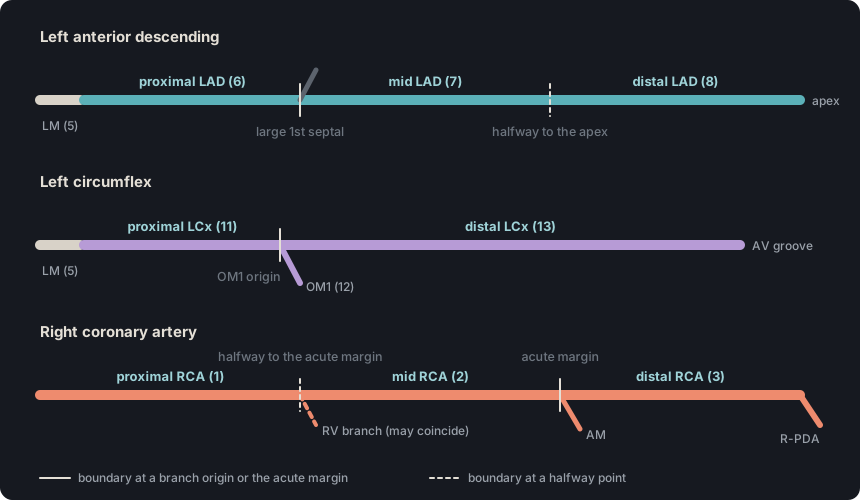
Segment boundaries are set by anatomic landmarks, not by thirds of length: in the LAD, the first large septal branch and half the distance to the apex; in the LCx, the origin of OM1; and in the RCA, the acute margin and half the distance to it.
Dominance is defined by the origin of the posterior descending artery, not by the size of the RCA, and it is stated in the findings.
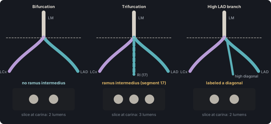
The ramus intermedius exists only when the left main trifurcates; if it bifurcates, a high-origin branch is named a diagonal or an obtuse marginal according to its vessel of origin.
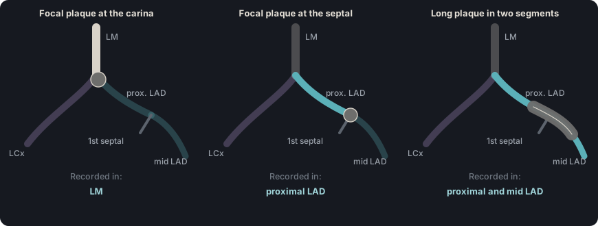
A focal lesion at the junction of two segments, including a bifurcation, is recorded only in the preceding, more proximal segment; a long plaque that spans both segments is recorded in both.
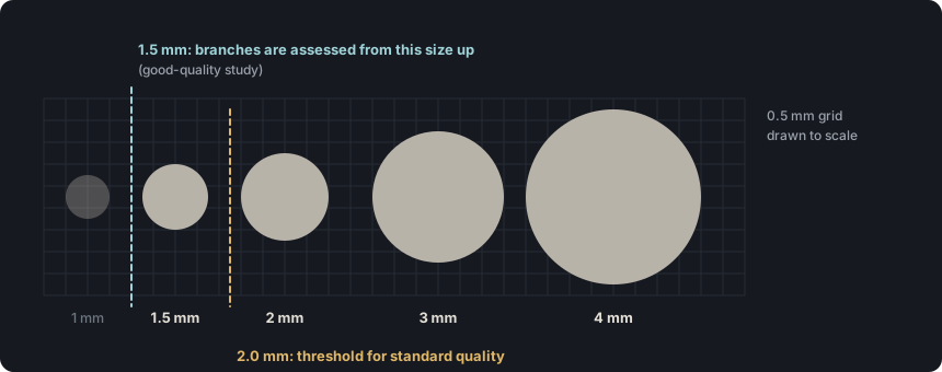
In a good-quality scan, all branches of 1.5 mm or more are assessed (2.0 mm or more if image quality is standard); if a segment of 1.5 mm or more cannot be interpreted, name it and state the reason.
Tap a segment or its number to see its name and boundary.
Figure 1. The map's coronary tree with the numbering of SCCT 2026 Table 1. The toggle shows which posterior branches exist in each dominance pattern. The ramus intermedius (17) is dashed because it exists only when the left main trifurcates, and the acute marginal (AM) has no number in the guideline.
Table 1. Coronary segments according to Table 1 of the SCCT 2026 consensus. The acute marginal is described in the guideline text (p. 12) and has no number.
No.
Abbreviation
Segment
Boundary per Table 1
1
pRCA
Proximal RCA
From the RCA ostium to one-half the distance to the acute margin of the right ventricle (may coincide with the origin of a right ventricular branch).
2
mRCA
Mid RCA
From the end of the proximal RCA to the acute margin of the right ventricle (may coincide with the origin of the acute marginal).
3
dRCA
Distal RCA
From the end of the mid RCA to the origin of the R-PDA (or to the interventricular groove if there is no R-PDA).
4
R-PDA
Right posterior descending artery (R-PDA)
Branch of the RCA following the posterior interventricular groove.
5
LM
Left main (LM)
From the LM ostium to its bifurcation into the LAD and LCx, or its trifurcation into the LAD, LCx and ramus intermedius.
6
pLAD
Proximal LAD
From the end of the LM to the first large septal branch.
7
mLAD
Mid LAD
From the end of the proximal LAD to one-half the distance to the apex.
8
dLAD
Distal LAD
From the end of the mid LAD to the end of the LAD.
9
D1
First diagonal (D1)
The first of the two largest diagonal branches traversing the anterolateral left ventricular wall.
10
D2
Second diagonal (D2)
The second of the two largest diagonal branches.
11
pLCx
Proximal LCx
From the end of the LM to the origin of OM1.
12
OM1
First obtuse marginal (OM1)
First branch of the LCx traversing the lateral wall of the left ventricle.
13
dLCx
Distal LCx ("LCx mid-distal" in the app)
Remainder of the LCx within the atrioventricular groove, distal to OM1.
14
OM2
Second obtuse marginal (OM2)
Second branch of the LCx traversing the lateral wall of the left ventricle.
15
L-PDA
Left posterior descending artery (L-PDA)
PDA arising from the LCx and following the posterior interventricular groove.
16
R-PL
Right posterolateral branch (R-PLB in the app)
Posterolateral branch of the RCA crossing the posterior interventricular groove.
17
RI
Ramus intermedius (RI)
Third vessel (trifurcation) arising from the LM between the LAD and LCx.
18
L-PL
Left posterolateral branch (L-PLB in the app)
Branch of the LCx traversing the posterolateral wall of the left ventricle.
Source: SCCT 2026, Table 1, p. 47 (adapted).
SCCT coronary artery segmentation model (18 segments)#
Also seen as:SCCT 18-segment model, 18-segment coronary model
The SCCT adapted the AHA segment model for interpreting coronary CT angiography. Its version has 18 segments. The ramus intermedius is segment 17 and the left posterolateral branch is segment 18.
In practice. Each segment is reviewed in long-axis and cross-sectional planes, and plaque type and stenosis grade are recorded for each one. The report uses the anatomic name, such as mid LAD, not the segment number.
Pitfall. Writing "segment 7 stenosis" in the report forces the referring physician to look up the diagram. The guideline recommends stating the anatomic location.
Other sources. The angiographic SYNTAX scheme also derives from the AHA model, but it uses 16 segments with subsegments, such as 9a or 12b, and numbers some branches differently: the ramus intermedius is 12, not 17. (Sianos 2005, SYNTAX, Table 1 and Fig. 1, pp. 220–221)
On the map. The map shows the 18 segments plus the acute marginal, and the posterior branches change with the selected dominance. The template names each segment by its anatomic location, never by its number.
Source: SCCT 2026, Tables 1 and 5, pp. 47 and 51 · «The standard American Heart Association (AHA) coronary artery segmentation model has been adapted for interpreting CCTA with minimal alterations»
Also seen as:LMCA, left main stem(British usage), LMS(British usage; interventional literature), left main trunk(Mostly Asian interventional literature), LMT(Mostly Asian interventional literature)
The left main is segment 5. It runs from its ostium to the bifurcation into the LAD and LCx, or to the trifurcation into the LAD, LCx and ramus intermedius.
In practice. It is reviewed in long-axis and cross-sectional planes from the ostium to the carina of the bifurcation. The guideline notes that a normal reference segment is hard to find in the left main. In that case, stenosis can be graded using normal reference diameters.
Pitfall. A bifurcation plaque does not belong to the ostial LAD. The preceding-segment rule assigns it to the LM, where a 50–99 % stenosis is CAD-RADS 4B.
Other sources. The interventional literature often divides the LM into ostial, shaft and distal (bifurcation) portions. SCCT 2026 treats it as a single segment and recommends stating the location in anatomic terms, not by number alone. (Dąbrowski 2022, section 2.3.5; SCCT 2026, p. 11)
On the map. The LM has its own segment on the map. If a 50–99 % stenosis is marked there, the template shows CAD-RADS 4B; with a total occlusion it shows CAD-RADS 5.
Source: SCCT 2026, Table 1, p. 47 · «Ostium of LM to the bifurcation of the LAD and LCx or trifurcation to LAD, LCx and ramus intermedius»
Proximal, mid and distal left anterior descending artery LAD · segments 6, 7 and 8#
Also seen as:pLAD, mLAD, dLAD, anterior interventricular artery(Anatomical name)
The proximal LAD runs from the end of the LM to the first large septal branch. The mid LAD continues to one-half the distance to the apex, and the distal LAD is the rest of the artery.
In practice. On the curved reconstruction of the LAD, find the first large septal branch, where the proximal segment ends. Then estimate the midpoint of the remaining course to the apex; that point separates the mid from the distal LAD.
Pitfall. Do not keep using the first diagonal as the end of the proximal LAD, as the 2014 guideline did. Table 1 of the 2026 guideline defines that boundary only by the first large septal branch.
Other sources. SCCT 2014 ended the proximal LAD at the first large septal branch or at D1, whichever arose first. This glossary follows SCCT 2026, which uses only the septal branch. (SCCT 2014, Appendix 1, p. 358)
On the map. The map divides the LAD into three segments. The first septal branch is drawn exactly where the proximal LAD ends, but it has no segment of its own. Open the example on the map (CAD-RADS 3/P1) →
How the map's template words it
Left anterior descending (LAD): mid segment: partially calcified plaque resulting in moderate stenosis (50-69%); the remainder is unremarkable. Branches D1, D2: no plaque.
Source: SCCT 2026, Table 1, p. 47 · «End of LM to the first large septal branch»
Figure 2. Where each segment ends, with the arteries straightened as in a curved reconstruction. A solid line marks a boundary at a branch origin or at the acute margin; a dashed line marks a boundary halfway along a course.
First and second diagonal branches D1 (9) · D2 (10)#
Also seen as:first diagonal artery, second diagonal artery
D1 and D2 are the two largest diagonal branches traversing the anterolateral wall of the left ventricle. Of the two, D1 arises first and D2 arises next.
In practice. Review every diagonal of 1.5 mm or more and pick the two largest. Of these, the more proximal one is D1. The guideline also asks that the number and size of branch vessels be described.
Pitfall. A small diagonal is not D1 just because it arises first. Table 1 reserves the names D1 and D2 for the two largest diagonals.
Other sources. SCCT 2014 and SYNTAX name as D1 the first diagonal to arise, not the first of the two largest. In SYNTAX, D2 arises from the distal LAD or at its junction with the mid LAD. (SCCT 2014, Appendix 1, p. 358; Sianos 2005, SYNTAX, Fig. 1, p. 221)
On the map. D1 and D2 each have their own segment on the map. A third diagonal has no segment of its own.
Source: SCCT 2026, Table 1, p. 47 · «First of the largest two diagonal branches traversing the anterolateral left ventricular wall»
Also seen as:ramus intermedius artery, ramus medianus, intermediate artery, intermediate branch, ramus(Report shorthand)
The ramus intermedius arises directly from the left main, between the LAD and the LCx, when the left main trifurcates instead of bifurcating. In SCCT numbering it is segment 17.
In practice. Before reporting a ramus intermedius, follow the branch back to its origin. If it arises from the LCx very close to the bifurcation, the ASCI 2024 consensus classifies it as the first obtuse marginal (OM1). When it arises from the LAD, it is reported as a high-origin diagonal.
Other sources. In the left main PCI literature, a major branch arising within 3 mm of the bifurcation is counted as part of a trifurcation, and some authors call it a ramus intermedius. The SCCT CCTA guidelines do not use that distance. (Tamburino 2009; Kovacevic 2021; ASCI 2024 consensus)
On the map. The RI has its own segment on the map and can be set to "Ramus intermedius absent". While it is absent, its findings are excluded from the template and the category. Open the example on the map (CAD-RADS 2/P1) →
Source: SCCT 2026, Table 1, p. 47 · «Third vessel (trifurcation) originating from the left main between the LAD and LCx»
Figure 3. The ramus intermedius is confirmed by counting lumens on a cross-section placed at the carina. If the left main bifurcates, a high branch from the LAD is a diagonal and a high branch from the LCx is an obtuse marginal.
Proximal and distal left circumflex artery LCx · segments 11 and 13#
Also seen as:Cx, circ(Informal), circumflex artery, pLCx, dLCx
The proximal LCx runs from the end of the LM to the origin of OM1. The distal LCx is the rest of the artery within the atrioventricular groove. The guideline does not define a mid LCx.
In practice. The only boundary is the origin of OM1, found on the curved reconstruction of the LCx. What lies before it is the proximal LCx, and what continues within the atrioventricular groove is the distal LCx.
Pitfall. The term "mid LCx" should not appear in the report. The guideline divides the circumflex only into proximal and distal.
Other sources. SCCT 2014 called segment 13 the "mid and distal LCx". In the SYNTAX scheme, the distal LCx begins after the origin of the last obtuse marginal, not OM1. This glossary follows SCCT 2026. (SCCT 2014, Appendix 1, p. 358; Sianos 2005, SYNTAX, Fig. 1, p. 221)
On the map. The map labels guideline segment 13 as "LCx mid-distal" and draws OM1 exactly where the proximal LCx ends. In the findings, the template calls it the distal segment of the circumflex. Open the example on the map (CAD-RADS 3/P1) →
How the map's template words it
Circumflex (LCx): proximal segment: partially calcified plaque causing moderate stenosis (50-69%); the rest of the vessel is unremarkable. Branches OM1, OM2: no plaque.
Source: SCCT 2026, Table 1, p. 47 · «Remainder of the LCx within the atrioventricular groove distal to the OM1»
First and second obtuse marginal branches OM1 (12) · OM2 (14)#
Also seen as:OM, obtuse marginal artery, OM branch, left marginal branch(Anatomical name), marginal branch of the circumflex
The obtuse marginals arise from the LCx and traverse the lateral wall of the left ventricle. The first is OM1, whose origin marks the end of the proximal LCx, and the second is OM2.
In practice. Follow them from the LCx to the lateral wall and number them by order of origin, unlike the diagonals, which are chosen by size. Find OM1 first, because its origin sets the boundary between the proximal and distal LCx.
Pitfall. The LCx branch that traverses the posterolateral wall is not an obtuse marginal. Table 1 defines it as the left posterolateral branch, segment 18.
Other sources. In the SYNTAX scheme, the obtuse marginals are segments 12a and 12b, and 14 is the left posterolateral branch. This glossary follows SCCT 2026, where OM2 is 14 and the left posterolateral branch is 18. (Sianos 2005, SYNTAX, Table 1 and Fig. 1, pp. 220–221)
On the map. OM1 and OM2 each have their own segment on the map; a third obtuse marginal does not.
Source: SCCT 2026, Table 1, p. 47 · «First branch from the LCX traversing the lateral wall of the left ventricle»
Proximal, mid and distal right coronary artery RCA · segments 1, 2 and 3#
Also seen as:pRCA, mRCA, dRCA
The proximal RCA runs from the ostium to one-half the distance to the acute margin of the right ventricle. The mid RCA ends at the acute margin, and the distal RCA reaches the origin of the R-PDA.
In practice. The landmark is the acute margin of the right ventricle. The midpoint of the course to that point separates the proximal from the mid RCA and may coincide with a right ventricular branch. If there is no R-PDA, the distal RCA ends at the interventricular groove.
Pitfall. The RCA is not divided into thirds of length. Table 1 sets its boundaries by the acute margin of the right ventricle and the origin of the R-PDA.
Other sources. The 28-segment CASS model, optional in SCCT 2014, numbers the proximal, mid and distal RCA the same way (1, 2 and 3), but segment 5 is the right posterior atrioventricular segment (RPAS) and the left main is 11. (SCCT 2014, section 5.3 and Table 5, CASS model)
Right coronary (RCA): mid segment: calcified plaque producing mild stenosis (25-49%); the rest of the vessel is unremarkable. Branches AM, R-PDA, R-PLB: no plaque.
Also seen as:acute marginal artery, right marginal branch(Anatomical name), right marginal artery(Anatomical name), ramus marginalis dexter(Latin anatomical name)
The acute marginal arises from the RCA between the end of the mid RCA and the acute margin of the heart. The guideline describes it in the text, but Table 1 does not assign it a number.
In practice. It is found at the acute margin of the right ventricle. As with other segments, plaque in it is described by its anatomic name. If the branch measures less than 1.5 mm and the plaque is clearly seen, describe it and note the small caliber.
Pitfall. A right ventricular branch from the proximal or mid RCA is not the acute marginal. The acute marginal arises between the end of the mid RCA and the acute margin.
Other sources. Anatomy atlases and textbooks call the acute marginal the right marginal branch of the right coronary artery (Latin ramus marginalis dexter). It is the same artery; this glossary uses the SCCT 2026 name. (IMAIOS e-Anatomy)
On the map. Although Table 1 does not number it, the map includes the acute marginal as its own segment (AM) and draws it at the end of the mid RCA. Open the example on the map (CAD-RADS 2/P1) →
How the map's template words it
Right coronary (RCA): no atherosclerotic disease. Acute marginal (AM): non-calcified plaque resulting in mild stenosis (25-49%); Branches R-PDA, R-PLB: no plaque.
Source: SCCT 2026, coronary anatomy text, p. 12 · «The acute marginal branch originates between the end of mid RCA and acute margin of the heart.»
Right and left posterior descending artery; right and left posterolateral branch R-PDA (4) · L-PDA (15) · R-PLB (16) · L-PLB (18)#
Also seen as:PDA, posterior interventricular artery(Anatomical name), PLB, PLV(Mostly invasive angiography literature), posterior left ventricular branch, posterolateral artery
The posterior descending artery (R-PDA from the RCA, L-PDA from the LCx) runs in the posterior interventricular groove. The R-PLB crosses that groove from the RCA, and the L-PLB traverses the posterolateral wall from the LCx.
In practice. Follow each branch back to its origin on the curved reconstruction, because the vessel of origin defines its name. For the posterior descending artery, the origin also determines dominance. In a co-dominant circulation with both an R-PDA and an L-PDA, each is assessed as its own segment.
Other sources. In the SYNTAX score, the left posterolateral branch is segment 14 and the right posterolateral branches (16) are 16a, 16b and 16c. In SCCT 2026, 14 is OM2 and the L-PL is 18. (Sianos 2005, SYNTAX, Fig. 1; SCCT 2026, Table 1)
On the map. The map uses the abbreviations R-PLB and L-PLB, equivalent to R-PL and L-PL in Table 1. Depending on the dominance button, it shows the right branches, the left branches, or both. Open the example on the map (CAD-RADS 3/P1) →
How the map's template words it
Circumflex (LCx): no plaque or stenosis. Left PDA (L-PDA): partially calcified plaque producing moderate stenosis (50-69%); Branches OM1, OM2, L-PLB: no plaque.
Right coronary (RCA): no atherosclerotic disease. Right PDA (R-PDA): non-calcified plaque producing mild stenosis (25-49%); Branches AM, R-PLB: no plaque.
Also seen as:coronary artery dominance, right-dominant circulation, left-dominant circulation, codominance
The origin of the posterior descending artery defines dominance. It is right if the PDA arises from the RCA (70–80 %), left if it arises from the LCx (5–10 %), and co-dominant if both contribute (10–20 %).
In practice. Follow the posterior descending artery along the posterior interventricular groove to its vessel of origin. Establish dominance before assigning lesions in the posterior branches. Table 6 gives a strong recommendation to state it in the findings.
Pitfall. Dominance is not defined by the size of the RCA or by the posterolateral branch. The guideline defines it only by the origin of the posterior descending artery.
Other sources. Some texts call the circulation balanced or co-dominant when the posterior descending artery arises from the RCA and the posterolateral branches from the LCx. The SYNTAX score allows only right or left dominance. This glossary follows SCCT 2026. (Halpern 2011, p. 52; Sianos 2005, SYNTAX)
On the map. The "Right", "Left" and "Balanced" buttons change which posterior segments exist, and the template states the dominance in the findings. If the change would hide segments that contain data, the app warns before hiding and clearing them. Open the example on the map (CAD-RADS 4A/P1) →
How the map's template words it
Circumflex (LCx): distal segment: partially calcified plaque producing severe stenosis (70-99%); the rest of the vessel is unremarkable. Left PDA (L-PDA): non-calcified plaque producing mild stenosis (25-49%); Branches OM1, OM2, L-PLB: no plaque.
Right coronary (RCA): no atherosclerotic disease. Branch AM: no plaque.
Impression Severe obstructive CAD. CAD-RADS 4A/P1.
Source: SCCT 2026, text and Table 6, pp. 11–12 and 52 · «The coronary artery dominance is described based on the origin of the posterior descending artery (PDA).»
Assignment of disease at segment intersections and bifurcations#
When a lesion lies at the junction of two segments, including bifurcations, it is assigned to the preceding segment, the more proximal one in the direction of flow.
In practice. If a plaque occupies the left main carina and extends into the LAD ostium, it is recorded in the LM. The guideline recommends describing it as a bifurcation plaque extending into the LAD. A plaque that runs through two segments is recorded in both.
Pitfall. A focal lesion at the junction of two segments is not recorded in both. The guideline assigns it to the preceding segment, and duplicating it inflates the SIS for no reason.
Other sources. In the cath lab, bifurcation lesions are usually described with the Medina classification: three digits, 1 or 0, according to whether the proximal main vessel, the distal main vessel and the side branch are involved. It does not replace assignment to the preceding segment. (Medina 2006)
On the map. The app does not reassign the lesion on its own. The radiologist marks it in the preceding segment, and each marked segment counts separately toward the SIS. Open the example on the map (CAD-RADS 4B/P1) →
How the map's template words it
Left main (LM): partially calcified plaque resulting in moderate stenosis (50-69%).
Impression High-risk obstructive CAD due to left main stenosis (≥50%). CAD-RADS 4B/P1.
Source: SCCT 2026, coronary anatomy text, p. 11 · «Disease at the intersection of segments (including bifurcations) is assigned to the preceding coronary segment.»
Figure 4. A focal plaque at the junction of two segments is recorded in the preceding segment; a long plaque that spans two segments is recorded in both and adds two points to the SIS.
The guideline asks for all branches with a diameter of 1.5 mm or more to be assessed when the scan is of good quality, and those of 2.0 mm or more when it is of standard quality.
In practice. Diameter is estimated in the short axis. When a segment of 1.5 mm or more cannot be interpreted, name it and explain why. In a small vessel with clearly visible plaque, describe the plaque and note the small caliber.
Pitfall. Calling a vessel smaller than 1.5 mm nondiagnostic changes the category for no reason. CAD-RADS N is reserved for segments larger than 1.5 mm.
Other sources. CAD-RADS 1.0 and 2.0 grade stenosis in vessels larger than 1.5 mm, without regard to scan quality. In the SYNTAX score, vessels smaller than 1.5 mm are not considered candidates for PCI or surgery. (CAD-RADS 1.0, Tables 2 and 3; CAD-RADS 2.0, Tables 4 and 5; Sianos 2005, SYNTAX, p. 223)
On the map. Each segment has an "N" (nondiagnostic) option. Reserve it for segments of 1.5 mm or more that cannot be interpreted.
Source: SCCT 2026, stenosis text, p. 15, and Table 5, p. 51 · «With a good-quality scan, all branches with diameter ≥ 1.5 mm and with a standard quality scan, all branches with diameter ≥ 2.0 mm should be included in the interpretation»
Figure 5. Diameters to scale on a 0.5 mm grid. In a good-quality scan, branches of 1.5 mm or more are assessed; if quality is standard, the threshold rises to 2.0 mm.
Self-assessment
Short questions to check the key points. The answer appears when you choose.
Tap the segment on the tree
1The RCA is large, but the posterior descending artery arises from the LCx and no RCA branch runs in the posterior interventricular groove. Which dominance is stated in the findings?
The guideline describes dominance by the origin of the posterior descending artery; the size of the RCA plays no role. If the PDA arises from the LCx and the RCA gives rise to none, dominance is left. Table 6 gives a strong recommendation to state it in the findings. See the term
2In a good-quality scan, a 1.2 mm diagonal branch cannot be interpreted because of motion artifact. All segments of 1.5 mm or more are diagnostic and the maximum stenosis is 30 %. Which category applies?
N is reserved for studies in which a segment larger than 1.5 mm cannot be interpreted; a 1.2 mm branch does not justify it. The category is assigned from the diagnostic segments, and a maximum stenosis of 30 % is CAD-RADS 2. In addition, the N modifier accompanies a category only when a diagnostic segment has a stenosis of 50 % or more. See the term
References
Rajiah PS, Alkadhi H, Andreini D, Bullock-Palmer RP, Chinnaiyan K, Chow B, et al. Interpretation and reporting of coronary computed tomographic angiography (2026 update): an expert consensus document of the Society of Cardiovascular Computed Tomography (SCCT). J Cardiovasc Comput Tomogr. 2026. In press. doi:10.1016/j.jcct.2026.08.014 Cited pages refer to the journal pre-proof PDF.
Cury RC, Leipsic J, Abbara S, Achenbach S, Berman D, Bittencourt M, et al. CAD-RADS 2.0 - 2022 Coronary Artery Disease-Reporting and Data System: an expert consensus document of the SCCT, ACC, ACR and NASCI. J Cardiovasc Comput Tomogr. 2022;16(6):536-57. doi:10.1016/j.jcct.2022.07.002
Koweek L, Achenbach S, Berman DS, Carr JJ, Cury RC, Ghoshhajra B, et al. Standardized medical terminology for cardiac computed tomography 2023 update: an expert consensus document of the SCCT, AAPM, ACR, NASCI and RSNA. J Cardiovasc Comput Tomogr. 2023;17(5):345-54. doi:10.1016/j.jcct.2023.06.002
Narula J, Chandrashekhar Y, Ahmadi A, Abbara S, Berman DS, Blankstein R, et al. SCCT 2021 expert consensus document on coronary computed tomographic angiography: a report of the Society of Cardiovascular Computed Tomography. J Cardiovasc Comput Tomogr. 2021;15(3):192-217. doi:10.1016/j.jcct.2020.11.001
Neves PO, Andrade J, Monção H. Coronary artery calcium score: current status. Radiol Bras. 2017;50(3):182-9.
Tamburino C, Tomasello SD, Capodanno D, Di Salvo ME, Marzà F, Galassi AR. Long-term follow-up after drug eluting stent implantation in left main trifurcations. EuroIntervention. 2009;5(4):432-7. doi:10.4244/eijv5i4a68
Kovacevic M, Burzotta F, Elharty S, Besis G, Aurigemma C, Romagnoli E, et al. Left main trifurcation and its percutaneous treatment: what is known so far? Circ Cardiovasc Interv. 2021;14(3):e009872. doi:10.1161/CIRCINTERVENTIONS.120.009872
Kim C, Park CH, Lee BY, et al. 2024 consensus statement on coronary stenosis and plaque evaluation in CT angiography from the Asian Society of Cardiovascular Imaging-Practical Tutorial (ASCI-PT). Korean J Radiol. 2024;25(4):331-42. doi:10.3348/kjr.2024.0112
Sources for the “Other sources” notes
Sianos G, Morel MA, Kappetein AP, et al. The SYNTAX Score: an angiographic tool grading the complexity of coronary artery disease. EuroIntervention. 2005;1(2):219-27.
Leipsic J, Abbara S, Achenbach S, Cury R, Earls JP, Mancini GBJ, et al. SCCT guidelines for the interpretation and reporting of coronary CT angiography: a report of the Society of Cardiovascular Computed Tomography Guidelines Committee. J Cardiovasc Comput Tomogr. 2014;8(5):342-58. doi:10.1016/j.jcct.2014.07.003
Dąbrowski EJ, Kożuch M, Dobrzycki S. Left main coronary artery disease: current management and future perspectives. J Clin Med. 2022;11(19):5745. doi:10.3390/jcm11195745
Halpern EJ. Clinical Cardiac CT: Anatomy and Function. 2nd ed. New York: Thieme; 2011.
Medina A, Suárez de Lezo J, Pan M. Una clasificación simple de las lesiones coronarias en bifurcación. Rev Esp Cardiol. 2006;59(2):183. doi:10.1157/13084649
IMAIOS e-Anatomy. Right marginal branch of the right coronary artery (ramus marginalis dexter).
Cury RC, Abbara S, Achenbach S, et al. CAD-RADS: Coronary Artery Disease – Reporting and Data System. J Cardiovasc Comput Tomogr. 2016;10(4):269-81. doi:10.1016/j.jcct.2016.04.005