David F. Cuevas Cueto, MD · Updated September 27, 2026 · Sources: SCCT 2026 and CAD-RADS 2.0
Plaque is described by its composition, by any high-risk features it shows and by the patient's total plaque burden. The definitions follow the SCCT 2026 consensus and CAD-RADS 2.0.
Key points
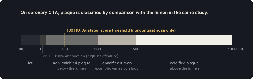
In calcified plaque, most of the plaque exceeds the attenuation of the contrast-enhanced lumen; non-calcified plaque stays below it, and partially calcified plaque has both components. The reference is the lumen of each study, not a fixed HU value.
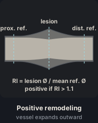
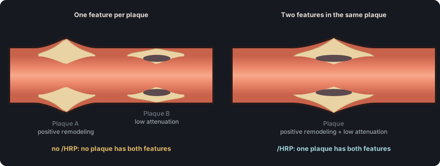
The /HRP modifier requires a single plaque to clearly show at least two of the four high-risk features: low attenuation, positive remodeling, spotty calcification and the napkin-ring sign.
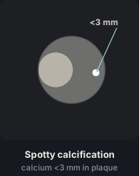
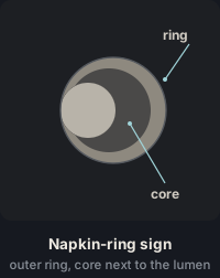
Low attenuation is defined as less than 30 HU, positive remodeling as a remodeling index greater than 1.1 and spotty calcification as a diameter less than 3 mm; the napkin-ring sign has no numeric threshold and is recognized by its shape on the cross-section.
The SIS counts segments with plaque, not plaques: an SIS of 1 to 2 corresponds to P1, 3 to 4 to P2, 5 to 7 to P3, and 8 or more to P4.
The Agatston score is calculated on the non-contrast phase; a score of 0 does not exclude non-calcified plaque, and when such plaque is present, burden is assigned with the SIS.
Plaque burden P1–P4 is independent of stenosis; when methods disagree, the more severe assessment is used, and no P is assigned in CAD-RADS 0.
Contrast-filled lumen
Non-calcified plaque
Low-attenuation core
Calcium
Peripheral ring
Pericoronary fat
Figure 1. Plaque types in cross-section, in CT grayscale. Calcified plaque exceeds the attenuation of the contrast-enhanced lumen, non-calcified plaque stays below it, and partially calcified plaque combines both components. Legend: contrast-enhanced lumen; non-calcified plaque; low-attenuation core; calcium; peripheral ring; pericoronary fat.
Figure 2. The four high-risk features. The /HRP modifier requires at least two in the same plaque. Positive remodeling is shown in a longitudinal view: the remodeling index is the outer vessel diameter at the lesion divided by the mean of the proximal and distal reference diameters, and it is positive when it exceeds 1.1.
Table 1. Coronary plaque burden classification. The Agatston score is used only if a non-contrast phase was acquired. If methods disagree, CAD-RADS 2.0 directs using the more severe assessment.
Plaque burden
CAD-RADS
Visual assessment
SIS (segments with plaque)
Agatston score
None
No P (CAD-RADS 0)
No plaque
0
0
Mild
P1
1–2 vessels with a mild amount of plaque
1–2
1–100
Moderate
P2
1–2 vessels with a moderate amount, or 3 vessels with a mild amount
3–4
101–300
Severe
P3
3 vessels with a moderate amount, or 1 vessel with a severe amount
5–7
301–999
Extensive
P4
2–3 vessels with a severe amount
≥8
≥1000
Source: SCCT 2026, Table 3, p. 49, based on CAD-RADS 2.0, Table 2. CAD-RADS 2.0 lists ">1000" as the P4 calcium score; SCCT 2026 lists "≥1000".
Also seen as:calcific plaque, hard plaque(Colloquial; not an SCCT term)
In calcified plaque, most of the plaque volume has higher attenuation than the opacified lumen. The criterion is relative to the vessel lumen, not a fixed HU threshold.
In practice. Assess it on cross-sections perpendicular to the vessel axis, with a wide window and, when available, a sharper kernel to reduce blooming. With dense calcium, report the highest visible grade and note that it may be overestimated.
Pitfall. The 130 HU Agatston threshold does not define calcified plaque on CTA. The reference is the attenuation of the opacified lumen.
Other sources. In semi-automated plaque quantification, most software classifies tissue above 350 HU as dense calcium. The glossary follows the SCCT 2026 visual criterion, which uses the contrast-enhanced lumen as the reference. (SCCT 2024 quantification, section 4.2)
On the map. Selecting «Calcified» in the editor disables the «Low attenuation» and «Napkin-ring sign» buttons. If the stenosis is 50–99 %, the template adds the caveat that the grade may be overestimated by calcium blooming. Open the example on the map (CAD-RADS 3/P1) →
How the map's template words it
Left anterior descending (LAD): proximal segment: calcified plaque causing moderate stenosis (50-69%), grade possibly overestimated by calcium blooming; the remainder is unremarkable. Branches D1, D2: no plaque.
Source: SCCT 2026, section on atherosclerotic plaque, p. 13 · «(largely) calcified plaque: attenuation values of most of the plaque exceed the values of the contrast-enhanced blood»
Figure 3. Plaque composition is judged against the lumen attenuation of each study. The 130 HU threshold belongs to the Agatston score, which is calculated on the non-contrast phase.
Also seen as:mixed plaque(Older term; discouraged by SCCT 2023), partially non-calcified plaque(Older term (SCCT 2014))
It combines a calcified and a non-calcified component. The SCCT uses this term rather than "mixed plaque". If one component makes up at least 75 %, it can be called predominantly calcified or predominantly non-calcified.
In practice. Review it on cross-sections and on multiplanar reformats, which help separate the non-calcified component from pericoronary fat. Plaque type is recorded for each segment; predominance is an optional qualifier in the text.
Pitfall. A plaque can be mistaken for calcified when beam hardening next to the calcium hides its non-calcified component. Review it with a wide window and in several planes.
Other sources. SCCT 2014 called it "partially non-calcified" and listed "predominantly calcified" and "predominantly non-calcified" separately, without a percentage threshold. Several articles call it "mixed plaque", a term SCCT 2024 discourages. (SCCT 2014, section 10.6.2; SCCT 2024 quantification, section 4.1)
On the map. In the editor it is marked with the «Partly calc.» button and accepts all four high-risk features. The template describes it as "partially calcified plaque".
Source: SCCT 2026, section on atherosclerotic plaque, p. 13 · «partially calcified: with both calcified and non-calcified components (Fig 6). A partially calcified plaque can be specified as predominantly calcified with ≥ 75 % of the plaque component calcified»
Also seen as:noncalcified plaque, NCP, soft plaque(Discouraged by SCCT), lipid-rich plaque(Discouraged by SCCT)
Non-calcified plaque has lower attenuation than the opacified lumen. The SCCT recommends this term instead of "soft" or "lipid-rich".
In practice. In addition to axial images, look for it on orthogonal and curved multiplanar reformats, because on axial images it can be missed or mistaken for a partial volume effect. It can also be confused with pericoronary fat. An unexplained change in caliber should prompt a closer look at the segment.
Pitfall. Calling it "soft" or "lipid" plaque is imprecise, because its attenuation depends on tube voltage and lumen opacification and does not predict histology in an individual patient.
Other sources. Quantification software subdivides non-calcified plaque by HU ranges with names such as fibrous, fibro-fatty or necrotic core. Thresholds and names vary between vendors, and SCCT 2024 discourages calling it "fibrous". (SCCT 2024 quantification, sections 4.1 and 4.2, Fig. 3)
On the map. It is marked with the «Noncalcified» button in the editor, and the template describes it as "non-calcified plaque" in its segment.
Source: SCCT 2026, section on atherosclerotic plaque, p. 13 · «non-calcified plaque: attenuation values of the plaque are below the values of the contrast-enhanced blood»
Also seen as:high-risk plaque features, vulnerable plaque(Older term (CAD-RADS 1.0)), vulnerable plaque features(Discouraged by SCCT 2023), adverse plaque features(Discouraged by SCCT 2023; in SCOT-HEART one feature was enough for 'adverse plaque')
A plaque is high-risk if it clearly shows at least two of four features (low attenuation, positive remodeling, spotty calcification and the napkin-ring sign). CAD-RADS 2.0 flags it with the /HRP modifier.
In practice. Review each plaque on cross-sections and count its features. If it has two or more, add /HRP to the category and name the features in the findings.
Pitfall. Features from different plaques are not added together to reach two. The modifier requires the features to be in the same plaque.
Other sources. CAD-RADS 1.0 used the "V" (vulnerability) modifier with the same two-or-more criterion; CAD-RADS 2.0 replaced it with "HRP". In SCOT-HEART, "adverse plaque" was defined by positive remodeling or low attenuation; one of them was enough. (CAD-RADS 1.0, section 3.3.4; CAD-RADS 2.0, section 3.3; Williams 2019, SCOT-HEART)
On the map. The editor for each segment lists the four features with an HRP n/4 counter. With two or more, the /HRP modifier is added; with only one, the template describes it as an isolated feature. Open the example on the map (CAD-RADS 2/P1/HRP) →
How the map's template words it
Left anterior descending (LAD): proximal segment: non-calcified plaque causing mild stenosis (25-49%) with high-risk plaque features (low attenuation and positive remodeling); the remainder is unremarkable. Branches D1, D2: no plaque.
Right coronary (RCA): mid segment: partially calcified plaque producing mild stenosis (25-49%) with spotty calcification as an isolated feature; the rest of the vessel is unremarkable. Branches AM, R-PDA, R-PLB: no plaque.
Source: CAD-RADS 2.0, section 3.3.4 and Figure 15 · «If a coronary plaque clearly demonstrates two or more high-risk features by CCTA, the modifier “HRP” (high risk plaque) should be added»
Figure 4. The /HRP modifier requires two features in the same plaque. Two plaques with one feature each do not trigger it; the features are described in the findings.
Also seen as:LAP, low-attenuation noncalcified plaque, low-density plaque
A non-calcified plaque has low attenuation, one of the high-risk features, when its internal attenuation is less than 30 HU. The measured value depends on tube voltage and lumen opacification.
In practice. Measure it on the cross-section with a small region of interest placed in the most hypodense part of the plaque, away from the lumen, calcium and pericoronary fat. Keep the acquisition tube voltage in mind.
Pitfall. Measuring next to dense calcium or at the border with pericoronary fat gives falsely low values, from beam hardening and partial volume.
Other sources. In volumetric quantification, the upper limit of the lowest-attenuation category ranges from 30 to 75 HU across vendors, and SCCT 2024 sets no absolute threshold. The glossary follows CAD-RADS 2.0. (SCCT 2024 quantification, section 4.2)
On the map. It is marked with the «Low attenuation» button in the editor, which is disabled when the plaque type is «Calcified».
Source: CAD-RADS 2.0, Figure 15; SCCT 2026, pp. 13–14 · «Low attenuation plaque, defined as non-calcified plaque with internal attenuation less than 30 HU.»
Also seen as:PR, positive remodelling, outward remodeling, expansive remodeling, compensatory enlargement(Older term (Glagov))
Positive remodeling is present when the outer vessel diameter at the plaque is more than 10 % larger than the reference diameter (remodeling index greater than 1.1). It is a high-risk feature.
In practice. On cross-sections, measure the outer vessel diameter, from outer wall border to outer wall border, at the plaque and at healthy proximal and distal reference segments. The index is the diameter at the lesion divided by the mean of the two reference diameters.
Pitfall. Using the lumen instead of the outer vessel contour to calculate the index is an error. Likewise, stenosis is graded against normal reference segments, not against the remodeled wall.
Other sources. The SCCT 2024 quantification document calculates the index with vessel areas, not diameters; it considers it positive at 1.10 or higher and negative below 0.90. The glossary uses diameters, as CAD-RADS 2.0 does. (SCCT 2024 quantification, Table 2)
On the map. It is marked with the «Positive remodeling» button in the editor, which remains available even when the plaque type is «Calcified».
Source: CAD-RADS 2.0, Figure 15; SCCT 2026, p. 13 · «Positive remodeling, defined as the ratio of outer vessel diameter at the site of plaque divided by the average outer diameter of the proximal and distal vessel greater than 1.1»
Spotty calcification is a calcium deposit less than 3 mm in diameter within a plaque. It is one of the high-risk features.
In practice. Measure the largest diameter of the deposit on the cross-section and the curved reformat, with a wide window to limit blooming. If it measures 3 mm or more, it no longer meets the criterion.
Pitfall. A small, isolated calcified plaque is not spotty calcification. The feature describes a calcium focus smaller than 3 mm within a plaque.
Other sources. Some authors define it relative to the vessel: its length is less than 1.5 times the vessel diameter and its width less than two-thirds of that diameter. The glossary follows the 3 mm limit of SCCT 2026. (van Rosendael 2024, Table 1; Yang 2021, Methods)
On the map. In the editor it is marked with the «Spotty calcification» button, and the template describes it as "spotty calcification".
Source: SCCT 2026, section on atherosclerotic plaque, p. 13 · «Features of high-risk plaque (HRP) include positive remodeling (outer vessel diameter >10% larger than the reference diameter), spotty calcification (diameter <3 mm)»
Also seen as:NRS, napkin ring sign, ring-like enhancement(Older term), ring-like sign
The napkin-ring sign is a high-risk feature seen on the cross-section of a non-calcified plaque: a low-attenuation center, apparently in contact with the lumen, surrounded by a ring of higher attenuation.
In practice. Look for it on cross-sections perpendicular to the vessel centerline, with a window setting that clearly separates the attenuation levels within the plaque. The ring is non-calcified tissue that is denser than the center; it is not calcium.
Pitfall. It can be mimicked by the calcified rim of a partially calcified plaque and by partial volume artifact at the plaque–lumen interface.
Other sources. Some reviews specify that the peripheral ring does not exceed 130 HU, and some studies call it ring-like enhancement. The 2024 SCCT quantification consensus states that the sign does not require measuring attenuation. (van Rosendael 2024, Table 1; Pozo 2016; SCCT 2024 quantification, section 4.1)
On the map. The editor records it with the «Napkin-ring sign» button, which is disabled when the plaque type is «Calcified».
Source: SCCT 2026, section on atherosclerotic plaque, p. 14 · «napkin-ring sign (central low attenuation apparently in contact with the lumen surrounded by ring-like higher-attenuation plaque tissue)»
Also seen as:CAD-RADS plaque burden, overall plaque burden
Plaque burden is the amount of coronary plaque, regardless of stenosis. CAD-RADS 2.0 expresses it as P1 (mild), P2 (moderate), P3 (severe) or P4 (extensive), based on the SIS, the Agatston score or visual assessment.
In practice. SCCT 2026 recommends estimating burden visually and combining that estimate with the Agatston score, if acquired, or with the SIS. When methods disagree, CAD-RADS 2.0 directs using the more severe assessment. No P is assigned in CAD-RADS 0.
Pitfall. With abundant non-calcified plaque, P should not be assigned from the Agatston score alone, because burden would be underestimated; P is adjusted upward.
Other sources. In CT plaque quantification, "plaque burden" also refers to the percentage of vessel area or volume occupied by plaque. The glossary uses it as CAD-RADS 2.0 does, for the patient's overall amount of plaque. (SCCT 2024 quantification, Table 2)
On the map. The P in the «Modifiers P · I» panel is taken from the map's SIS, from an Agatston score you enter, or from a manual choice. The template prints "Plaque burden: mild (P1; SIS 2)." Open the example on the map (CAD-RADS 2/P3) →
How the map's template words it
Left main (LM): calcified plaque resulting in minimal stenosis (1-24%).
Left anterior descending (LAD): proximal segment: calcified plaque causing minimal stenosis (1-24%); mid segment: partially calcified plaque resulting in mild stenosis (25-49%); the remainder is unremarkable. Branches D1, D2: no plaque.
Circumflex (LCx): proximal segment: calcified plaque causing minimal stenosis (1-24%); the rest of the vessel is unremarkable. Branches OM1, OM2: no plaque.
Right coronary (RCA): proximal segment: calcified plaque causing minimal stenosis (1-24%); the rest of the vessel is unremarkable. Branches AM, R-PDA, R-PLB: no plaque.
Source: SCCT 2026, text and Table 3, pp. 14 and 49; CAD-RADS 2.0, section 3.2.1 · «CAD-RADS 2.0 introduces the P1 to P4 descriptors which are used to denote increasing categories of plaque burden (P1=mild, P2=moderate, P3=severe and P4=extensive)»
The SIS counts coronary segments with plaque, regardless of plaque composition or stenosis grade. Each segment with plaque scores 1 point, and the maximum is 16. It is more reproducible than visual estimation of burden.
In practice. Go through the coronary tree segment by segment and count each one that has plaque, even if minimal. If plaque sits at the junction of two segments, including bifurcations, it is assigned to the preceding segment.
Pitfall. Counting plaques instead of segments is an error. A long plaque in the proximal and mid LAD scores 2 points, and three plaques in one segment score 1.
Other sources. Some studies call disease with plaque in more than 4 segments "extensive". The glossary follows CAD-RADS 2.0, in which 5 to 7 segments correspond to P3 (severe) and only 8 or more to P4 (extensive). (Bittencourt 2014; SCCT 2026, Table 3)
On the map. In «Auto (map)» mode, the app counts the segments where plaque was marked, excludes those marked N (nondiagnostic) and converts the total to P1–P4. Open the example on the map (CAD-RADS 2/P2) →
How the map's template words it
Left anterior descending (LAD): proximal segment: non-calcified plaque causing mild stenosis (25-49%); mid segment: non-calcified plaque resulting in mild stenosis (25-49%); the remainder is unremarkable. Branches D1, D2: no plaque.
Right coronary (RCA): mid segment: calcified plaque producing minimal stenosis (1-24%); the rest of the vessel is unremarkable. Branches AM, R-PDA, R-PLB: no plaque.
Source: SCCT 2026, section on plaque volume, p. 14 · «A score of 1 is given for each segment with plaque, with the highest possible score of 16.»
SIS 0No plaque
Figure 5. SIS counter. Tap each segment that has plaque, of any composition or grade; the counter adds one point per segment and shows the corresponding plaque burden category. Tap a segment again to remove it.
Also seen as:calcium score(Discouraged by SCCT 2023), CAC score(Discouraged by SCCT 2023), coronary artery calcium score(Long form of CAC score, discouraged by SCCT 2023), CACS(Abbreviation of CAC score, discouraged by SCCT 2023), Agatston calcium score
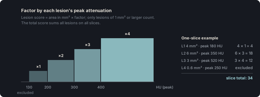
For each coronary lesion, the area of calcium above the 130 HU threshold is multiplied by a factor of 1 to 4 according to its peak attenuation. The score is the sum of these products.
In practice. It is calculated on the non-contrast ECG-gated acquisition, before the angiogram, with the standard protocol of 120 kVp and 3 mm slices. If performed, the total is reported and can be used to assign plaque burden.
Pitfall. A score of 0 does not mean absence of plaque. Non-calcified plaque adds no calcium, and when it is present, burden is assigned with the SIS.
Other sources. The SCCT CAC-DRS (2018) groups the score into A0 (0), A1 (1–99), A2 (100–299) and A3 (300 or more), and adds the number of arteries with calcium (for example, A2/N3). Its cutoffs differ slightly from those of P1–P4. (CAC-DRS, Hecht 2018; Obisesan 2021; SCCT 2026, Table 3)
On the map. In «Calcium (Agatston)» mode you enter the value and the app converts it to P1–P4. The template prints, for example, "Plaque burden: moderate (P2; Agatston 260)."
Source: Neves PO, et al. Radiol Bras 2017 (review), section "Agatston method" · «The Agatston method uses the weighted sum of lesions with a density above 130 HU, multiplying the area of calcium by a factor related to maximum plaque attenuation»
Figure 6. The Agatston score multiplies the area of each lesion by a factor that depends on its peak attenuation and sums the results for all lesions on all slices.
Self-assessment
Short questions to check the key points. The answer appears when you choose.
1A continuous plaque runs through the proximal, mid and distal RCA. There is also a plaque in the proximal LAD and another in the proximal LCx, and no other plaque. What is the SIS and what plaque burden does it indicate?
The SIS counts segments, not plaques. The continuous plaque scores 3 points, one for each RCA segment; with the proximal LAD and proximal LCx the total is 5, which corresponds to P3. See the term
2In the proximal LAD there is a plaque with positive remodeling and no other feature. In the mid LAD, separate from the first, another non-calcified plaque has an internal attenuation of 20 HU and no other feature. How is this reflected in the CAD-RADS category?
The modifier requires two or more features in the same plaque. Here each plaque has one: the features are described in the findings, but the category does not carry /HRP. See the term
3The Agatston score is 0. On CTA there is non-calcified plaque in the proximal LAD, mid LAD, proximal LCx and proximal RCA, with no stenosis of 50 % or more, and no other plaque. What plaque burden is assigned?
When methods disagree, the more severe assessment is used. The Agatston score does not measure non-calcified plaque; the SIS is 4, which corresponds to P2. See the term
4In the proximal LAD, the outer vessel diameter at the plaque is 4.0 mm and the lumen diameter is 2.6 mm. The outer reference diameters are 3.9 mm in the healthy proximal segment and 3.1 mm in the distal segment. What is the remodeling index?
Divide the outer diameter at the lesion by the mean of the references: 4.0 / 3.5 = 1.14, greater than 1.1. Using only the proximal reference gives 1.03, and using the lumen gives 0.74. See the term
5On the cross-section of a proximal RCA plaque there is a large low-attenuation center in contact with the lumen, surrounded by a thin, continuous ring of dense calcium. How is it described?
In the napkin-ring sign, the ring is non-calcified plaque tissue denser than the center, and CAD-RADS 2.0 defines it in a non-calcified plaque. A ring of dense calcium does not meet the criterion and, together with the non-calcified center, makes the plaque partially calcified. See the term
6On the non-contrast phase, the only coronary calcium deposit is in the proximal LAD and appears on a single slice: it measures 5 mm² and its peak attenuation is 320 HU. CTA shows no other plaque. What is the Agatston score and what plaque burden does it indicate?
The factor depends on the lesion's peak attenuation: 300 to 399 HU gives 3, so the score is 5 × 3 = 15. A value of 1 to 100 corresponds to P1, as does an SIS of 1. See the term
References
Rajiah PS, Alkadhi H, Andreini D, Bullock-Palmer RP, Chinnaiyan K, Chow B, et al. Interpretation and reporting of coronary computed tomographic angiography (2026 update): an expert consensus document of the Society of Cardiovascular Computed Tomography (SCCT). J Cardiovasc Comput Tomogr. 2026. In press. doi:10.1016/j.jcct.2026.08.014 Cited pages refer to the journal pre-proof PDF.
Cury RC, Leipsic J, Abbara S, Achenbach S, Berman D, Bittencourt M, et al. CAD-RADS 2.0 - 2022 Coronary Artery Disease-Reporting and Data System: an expert consensus document of the SCCT, ACC, ACR and NASCI. J Cardiovasc Comput Tomogr. 2022;16(6):536-57. doi:10.1016/j.jcct.2022.07.002
Koweek L, Achenbach S, Berman DS, Carr JJ, Cury RC, Ghoshhajra B, et al. Standardized medical terminology for cardiac computed tomography 2023 update: an expert consensus document of the SCCT, AAPM, ACR, NASCI and RSNA. J Cardiovasc Comput Tomogr. 2023;17(5):345-54. doi:10.1016/j.jcct.2023.06.002
Narula J, Chandrashekhar Y, Ahmadi A, Abbara S, Berman DS, Blankstein R, et al. SCCT 2021 expert consensus document on coronary computed tomographic angiography: a report of the Society of Cardiovascular Computed Tomography. J Cardiovasc Comput Tomogr. 2021;15(3):192-217. doi:10.1016/j.jcct.2020.11.001
Neves PO, Andrade J, Monção H. Coronary artery calcium score: current status. Radiol Bras. 2017;50(3):182-9.
Tamburino C, Tomasello SD, Capodanno D, Di Salvo ME, Marzà F, Galassi AR. Long-term follow-up after drug eluting stent implantation in left main trifurcations. EuroIntervention. 2009;5(4):432-7. doi:10.4244/eijv5i4a68
Kovacevic M, Burzotta F, Elharty S, Besis G, Aurigemma C, Romagnoli E, et al. Left main trifurcation and its percutaneous treatment: what is known so far? Circ Cardiovasc Interv. 2021;14(3):e009872. doi:10.1161/CIRCINTERVENTIONS.120.009872
Kim C, Park CH, Lee BY, et al. 2024 consensus statement on coronary stenosis and plaque evaluation in CT angiography from the Asian Society of Cardiovascular Imaging-Practical Tutorial (ASCI-PT). Korean J Radiol. 2024;25(4):331-42. doi:10.3348/kjr.2024.0112
Sources for the “Other sources” notes
Leipsic J, Abbara S, Achenbach S, Cury R, Earls JP, Mancini GBJ, et al. SCCT guidelines for the interpretation and reporting of coronary CT angiography: a report of the Society of Cardiovascular Computed Tomography Guidelines Committee. J Cardiovasc Comput Tomogr. 2014;8(5):342-58. doi:10.1016/j.jcct.2014.07.003
Cury RC, Abbara S, Achenbach S, et al. CAD-RADS: Coronary Artery Disease – Reporting and Data System. J Cardiovasc Comput Tomogr. 2016;10(4):269-81. doi:10.1016/j.jcct.2016.04.005
Nieman K, García-García HM, Hideo-Kajita A, et al. Standards for quantitative assessments by coronary computed tomography angiography (CCTA): an expert consensus document of the SCCT. J Cardiovasc Comput Tomogr. 2024;18(5):429-43. doi:10.1016/j.jcct.2024.05.232
Williams MC, Moss AJ, Dweck M, et al. Coronary artery plaque characteristics associated with adverse outcomes in the SCOT-HEART study. J Am Coll Cardiol. 2019;73(3):291-301. doi:10.1016/j.jacc.2018.10.066
van Rosendael SE, Shiyovich A, Cardoso RN, Souza Freire CV, van Rosendael AR, Lin FY, et al. The role of cardiac computed tomography angiography in risk stratification for coronary artery disease. J Soc Cardiovasc Angiogr Interv. 2024;3(11):102230. doi:10.1016/j.jscai.2024.102230
Yang S, Lee JM, Hoshino M, Murai T, Choi KH, Hwang D, et al. Prognostic implications of comprehensive whole vessel plaque quantification using coronary computed tomography angiography. JACC Asia. 2021;1(1):37-48. doi:10.1016/j.jacasi.2021.05.003
Pozo E, Agudo-Quilez P, Rojas-González A, Alvarado T, Olivera MJ, Jiménez-Borreguero LJ, et al. Noninvasive diagnosis of vulnerable coronary plaque. World J Cardiol. 2016;8(9):520-33. doi:10.4330/wjc.v8.i9.520
Bittencourt MS, Hulten E, Ghoshhajra B, et al. Prognostic value of nonobstructive and obstructive coronary artery disease detected by coronary computed tomography angiography to identify cardiovascular events. Circ Cardiovasc Imaging. 2014;7(2):282-91. doi:10.1161/CIRCIMAGING.113.001047
Hecht HS, Blaha MJ, Kazerooni EA, et al. CAC-DRS: Coronary Artery Calcium Data and Reporting System. An expert consensus document of the Society of Cardiovascular Computed Tomography (SCCT). J Cardiovasc Comput Tomogr. 2018;12(3):185-91. doi:10.1016/j.jcct.2018.03.008
Obisesan OH, Osei AD, Uddin SMI, Dzaye O, Blaha MJ. An update on coronary artery calcium interpretation at chest and cardiac CT. Radiol Cardiothorac Imaging. 2021;3(1):e200484. doi:10.1148/ryct.2021200484