Coronary Stents and Bypass Grafts on CCTA (CAD-RADS 2.0)
David F. Cuevas Cueto, MD · Updated September 27, 2026 · Sources: SCCT 2026 and CAD-RADS 2.0
In a revascularized patient, each stent and each graft is described by its location and patency, and its stenosis is graded on the same scale as native vessels. The definitions follow the SCCT 2026 consensus and CAD-RADS 2.0, which flags stents and grafts with the modifiers S and G and leaves out of the category any stenosis bypassed by a fully patent graft.
Key points
A stent is patent only if contrast opacifies its lumen, because distal contrast can arrive through collaterals.
In-stent restenosis is assessed along the entire stent and at its edges, and it is graded on the same scale as native stenoses.
A non-evaluable stent gives CAD-RADS N if there is no other stenosis of 50 % or more, and S is kept, as in N/P2/S.
S and G indicate that stents or grafts are present, do not change the category, and are written in that order, after N, HRP and I.
A stenosis bypassed by a fully patent graft does not count toward the category, but the graft, its distal anastomosis and the distal runoff are graded.
An occluded graft gives CAD-RADS 5, and the native branches proximal to its anastomosis are interpreted again.
Table 1. How stents and grafts count toward the CAD-RADS 2.0 category. Stenosis in a stent or a graft is graded on the native vessel scale, and the patient's category comes from the most severe stenosis that counts.
Situation
How it counts toward the category
Modifier
CAD-RADS 2.0 example
Patent stent
Adds no grade. The category comes from the other lesions.
S
2/S
In-stent restenosis
Graded like a native stenosis (70–99 % gives 4A)
S
4A/P3/S
Occluded stent
100 %, category 5
S
—
Non-evaluable stent
N replaces the number if there is no other stenosis ≥50 %. If there is, N is added as a modifier.
S
N/P2/S
Patent graft
Adds no grade, and the native stenosis it bypasses does not count
G
2/P2/G
Graft stenosis
Graded like a native vessel, at an anastomosis or in the graft body
G
—
Occluded graft
100 %, category 5. Native branches proximal to the anastomosis are interpreted again.
G
5/P3/G
Native segment bypassed by a patent graft
Does not count. It is described in the findings.
G (for the graft)
2/P4/G
Source: CAD-RADS 2.0, sections 3.3.2, 3.3.3 and 3.3 (examples of modifier order), Figures 13 and 14; SCCT 2026, pp. 19–20 and Table 5, p. 51.
Also seen as:stent evaluability, non-assessable stent, nonevaluable stent
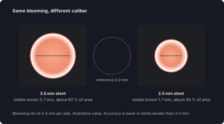
A stent is evaluable when its lumen is seen clearly enough to grade restenosis. If strut blooming, calcium or motion obscures the lumen, the stent is reported as non-evaluable.
In practice. Review it in cross-sectional and curved planar reformations with a wide window, thin sections and a sharp kernel, or with high-energy virtual monoenergetic images when available. Accuracy is lower in stents smaller than 3.0 mm.
Pitfall. Reporting a small stent as patent when blooming hides its lumen. If the lumen cannot be seen, the stent is non-evaluable and is reported as such.
Other sources. The 2010 appropriate use criteria rated CCTA as inappropriate for evaluating stents smaller than 3 mm or of unknown diameter. SCCT 2026 sets no minimum diameter and ties evaluation to the caliber, material and strut thickness of each stent. (Taylor 2010, cardiac CT AUC, Table 6; SCCT 2026, p. 19)
On the map. In the editor of a segment with a stent, the «N» button marks the stent as non-evaluable. The template writes "stent nonevaluable for restenosis due to artifact or small caliber" and keeps S. Open the example on the map (CAD-RADS N/P2/S) →
How the map's template words it
Left anterior descending (LAD): proximal segment: stent nonevaluable for restenosis due to artifact or small caliber; the remainder is unremarkable. Branches D1, D2: no plaque.
Circumflex (LCx): proximal segment: partially calcified plaque causing mild stenosis (25-49%); the rest of the vessel is unremarkable. Branches OM1, OM2: no plaque.
Right coronary (RCA): proximal segment: partially calcified plaque causing minimal stenosis (1-24%); mid segment: partially calcified plaque producing mild stenosis (25-49%); the rest of the vessel is unremarkable. Branches AM, R-PDA, R-PLB: no plaque.
Impression Nondiagnostic study for obstructive CAD. CAD-RADS N/P2/S.
Source: SCCT 2026, text on coronary stents (section h), blooming artifact (section e) and Table 5, pp. 19, 27 and 51 · «evaluation of in-stent restenosis (ISR) is highly dependent on stent size, material composition, strut thickness, overlapping segments, overlying dense calcification, and CCTA acquisition and reconstruction protocol»
Figure 1. The same band of blooming covers a larger fraction of the lumen in a small stent. Its thickness is illustrative and depends on the stent, the kernel and the scanner.
Also seen as:stent restenosis, in-stent stenosis, instent restenosis
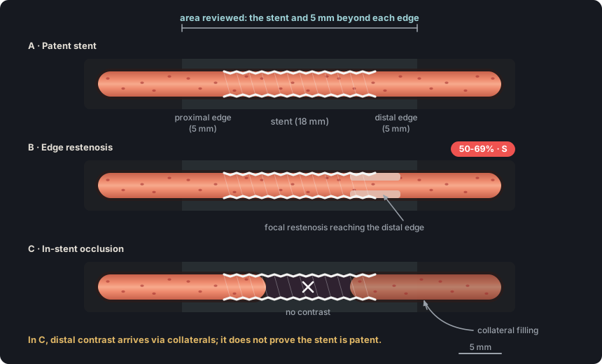
In-stent restenosis is narrowing of the lumen inside the stent or at its edges. SCCT 2026 classifies it as absent, mild (<50 %), significant (≥50 %), occlusion or non-evaluable.
In practice. Review the entire stent and 5 mm beyond each edge in cross-sectional and curved planar reformations. The grade is assigned on the native vessel scale, and the lesion is noted as focal or diffuse.
Pitfall. Mistaking the hypodense band that beam hardening leaves next to the struts for neointimal hyperplasia. Review another reconstructed phase before grading restenosis.
Other sources. Invasive angiography uses the Mehran classification, with four patterns. Pattern I is focal, 10 mm or shorter, and pattern II is diffuse but confined to the stent. Pattern III is diffuse and extends beyond the stent edges, and pattern IV is total occlusion. SCCT 2026 states that it can be adapted to CT. (Mehran 1999, Circulation, abstract; SCCT 2026, p. 19)
On the map. With the «▢ Stent» type, the grade set afterward is the restenosis. The template describes it as mild (<50%) or significant with its range, and the category treats it as a native stenosis. Open the example on the map (CAD-RADS 3/P1/S) →
How the map's template words it
Left anterior descending (LAD): proximal segment: significant in-stent restenosis (50-69%); the remainder is unremarkable. Branches D1, D2: no plaque.
Right coronary (RCA): mid segment: partially calcified plaque producing mild stenosis (25-49%); the rest of the vessel is unremarkable. Branches AM, R-PDA, R-PLB: no plaque.
Source: SCCT 2026, Table 5, interpretation consensus statements (strong recommendation), p. 51 · «In-stent restenosis is classified as absent, mild (<50%), significant (≥50%), occlusion, or non-evaluable.»
Figure 2. Restenosis is assessed along the entire stent and 5 mm beyond each edge. If the stent lumen does not opacify, distal contrast does not prove patency.
Also seen as:in-stent occlusion, stent occlusion, occluded stent
In-stent total occlusion is present when a segment of the stent lumen shows no contrast. It is equivalent to a 100 % stenosis and gives CAD-RADS 5.
In practice. Follow the lumen on curved planar reformation along the entire stent. The stent is patent if there is contrast proximal to, within and distal to it. The occlusion is reported as total, without assuming it is chronic.
Pitfall. Calling a stent patent because the distal artery opacifies. Distal contrast can arrive through collaterals, and patency requires contrast inside the stent.
Other sources. In the interventional literature, chronic total occlusion, including in-stent occlusion, is a 100 % stenosis with TIMI 0 flow for more than three months. Because CT cannot establish that duration, the glossary uses "in-stent total occlusion" and does not call it chronic. (Galassi 2019, EuroCTO consensus, Definition and epidemiology)
On the map. With the «▢ Stent» type at 100 %, the template writes "total in-stent occlusion (occluded stent)", the category is 5 and the impression adds a line about the occluded stent. Open the example on the map (CAD-RADS 5/P1/S) →
How the map's template words it
Right coronary (RCA): mid segment: total in-stent occlusion (occluded stent); the rest of the vessel is unremarkable. Branches AM, R-PDA, R-PLB: no plaque.
Impression Total occlusion (100%) of at least one vessel. CAD-RADS 5/P1/S.
Source: SCCT 2026, text on coronary stents (section h), p. 19 · «The presence of contrast medium distal to a stent alone is not a definitive sign of patency; contrast opacification should be demonstrated within the stent to establish patency.»
S indicates the presence of at least one coronary stent anywhere in the coronary tree. It does not change the category, which is assigned from the most severe stenosis, whether or not it lies within a stent.
In practice. A patent stent adds no grade. Restenosis is graded as a native stenosis, an occluded stent gives 5, and a non-evaluable stent gives N if there is no other stenosis of 50 % or more.
Pitfall. Omitting S because the stent is patent. S indicates the presence of the stent and is written even when there is no restenosis.
Other sources. CAD-RADS 1.0 placed S second, after N, and used V for vulnerable plaque. A patient with a stent and a moderate stenosis with high-risk plaque was coded "3/S/V". CAD-RADS 2.0 replaced V with HRP, added P and places S after HRP and I, as in "3/P3/HRP/S". (CAD-RADS 1.0, section 3.3.5, p. 275; CAD-RADS 2.0, sections 3.3.4 and 3.3)
On the map. The app adds S as soon as a segment has a «▢ Stent» lesion. That segment does not accept HRP features, and a stent with no other plaque gives category 1, a convention specific to the app. Open the example on the map (CAD-RADS 2/P1/S) →
How the map's template words it
Left anterior descending (LAD): proximal segment: patent stent, no significant restenosis; the remainder is unremarkable. Branches D1, D2: no plaque.
Circumflex (LCx): proximal segment: partially calcified plaque causing mild stenosis (25-49%); the rest of the vessel is unremarkable. Branches OM1, OM2: no plaque.
Right coronary (RCA): mid segment: partially calcified plaque producing mild stenosis (25-49%); the rest of the vessel is unremarkable. Branches AM, R-PDA, R-PLB: no plaque.
Source: CAD-RADS 2.0, section 3.3.2 · «The modifier “S” indicates the presence of at least one coronary stent anywhere in the coronary system.»
Arterial graft (internal mammary and radial artery) LIMA#
Also seen as:LITA(Current anatomical name (left internal thoracic artery)), internal thoracic artery graft, IMA graft, RIMA, radial artery graft, arterial conduit
An arterial graft uses one of the patient's arteries as the conduit. The left internal mammary artery is usually left attached to the subclavian artery and anastomosed to the LAD. The radial artery is a free graft.
In practice. Follow the internal mammary graft from the subclavian artery, behind the anterior chest wall, to its anastomosis with the LAD. The metal clips along its course cause beam-hardening artifact, which is reduced with iterative reconstruction or high-energy virtual monoenergetic images.
Pitfall. Mistaking the dark band left by metal clips next to the internal mammary graft for a stenosis. Confirm any stenosis with a wide window and in several planes.
Other sources. Current anatomical nomenclature and many surgical articles call the internal mammary artery the internal thoracic artery, abbreviated ITA. It is the same vessel. The glossary keeps "internal mammary" because it is the SCCT 2026 term, abbreviated LIMA. (IMAIOS e-Anatomy; Taggart 2016, NEJM; SCCT 2026, p. 5)
On the map. The «Grafts (bypass) ▾» menu includes LIMA-LAD and Radial-OM2, the arterial grafts, drawn as solid lines. The template names the conduit "left internal mammary artery" or "radial artery".
Source: SCCT 2026, text on coronary artery bypass grafts (section i) and beam-hardening artifact (section d), pp. 19 and 27 · «Coronary artery grafts are described by graft type (i.e., arterial or venous), proximal and distal coronary anastomosis sites and distal runoff.»
Also seen as:vein graft, venous graft, saphenous vein bypass graft, aortocoronary vein graft, long saphenous vein graft(Older term)
A vein graft is a segment of the great saphenous vein used as a free graft from the ascending aorta to the coronary artery. When it supplies several branches in series, it is called a sequential graft.
In practice. Count the grafts and look for each proximal anastomosis on the ascending aorta. In a sequential graft, review each segment, because the occlusion may be limited to one of them.
Pitfall. Missing an occluded graft because it does not opacify. An aortic stump or clips without an opacified graft suggest an occluded graft, and the finding is checked against the operative report.
Other sources. Other texts call the great saphenous vein the internal or long saphenous vein. All three names refer to the same vein, and the glossary uses "great", the term recommended in 2001 by an international phlebology consensus. (Chen 2009, Australas J Ultrasound Med)
On the map. The SVG-RCA, SVG-OM, SVG-D1, SVG-D2 and SVG-RI buttons draw saphenous vein grafts as dashed lines. The template describes them in a separate paragraph, before the native vessels. Open the example on the map (CAD-RADS 4A/P1/G) →
How the map's template words it
Coronary bypass grafts (CABG). The following grafts are assessed:
saphenous vein graft to the OM1: patent with 75% stenosis (body) (SVG-OM).
Circumflex (LCx): proximal segment: partially calcified plaque causing mild stenosis (25-49%); the rest of the vessel is unremarkable. Branches OM1, OM2: no plaque.
Impression Severe obstructive CAD. CAD-RADS 4A/P1/G.
Source: SCCT 2026, text on coronary artery bypass grafts (section i), pp. 19–20 · «High-density surgical material in the aortic root without adjacent opacified grafts (stump) or random vascular clips around the heart may indicate the presence of a completely occluded graft.»
Proximal and distal anastomoses, and distal runoff#
Also seen as:distal run-off, graft anastomosis
The proximal anastomosis joins the graft to its inflow vessel, and the distal anastomosis joins it to the recipient coronary artery. The distal runoff is the native coronary artery supplied by the graft beyond the distal anastomosis.
In practice. Document the patency of both anastomoses and describe the distal runoff, including its caliber and any stenoses. Lesions just beyond the distal anastomosis are easily missed and are sought with thin-slab MIP.
Pitfall. Excluding a stenosis in the distal runoff from the category as if it were bypassed. The anastomosis and the coronary artery distal to it are graded.
Other sources. In the surgical literature, the distal anastomosis is end-to-side when the end of the graft is joined to the side of the coronary artery. In a sequential graft, the intermediate anastomoses are side-to-side. The glossary calls both distal. (Tector 1984, Circulation, abstract)
On the map. The graft editor places its stenosis at «Proximal», «Body» or «Distal». The app excludes all target segments of a patent graft from the category, including the distal runoff, which the guideline does grade.
Source: SCCT 2026, Table 6, report components (strong recommendation), p. 52 · «Bypass graft (if present) to include type, location, patency, and distal runoff»
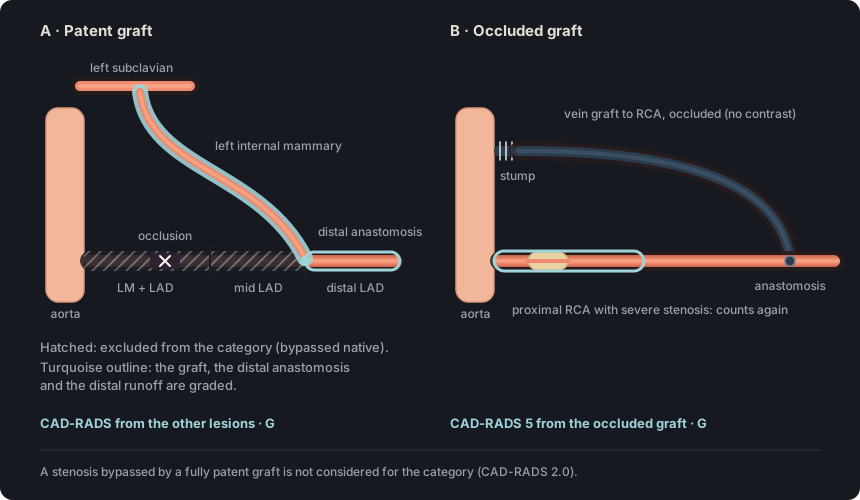
A native segment is bypassed when it lies proximal to the distal anastomosis of a graft. According to CAD-RADS 2.0, a stenosis bypassed by a fully patent graft is not considered for the category.
In practice. The bypassed stenosis is described in the findings, often as an expected occlusion of the native vessel. In that territory, the category takes into account the graft, its distal anastomosis and the coronary artery distal to it.
Pitfall. Continuing to exclude the native vessel when its graft is occluded. If the graft occludes, the native branches proximal to the anastomosis are interpreted and count toward the category.
On the map. While graft stenosis is below 50 %, the app excludes its target segments from the category and labels them "(revascularized by LIMA-LAD)". The impression lists them as "Native stenoses bypassed by a patent graft". Open the example on the map (CAD-RADS 2/P2/G) →
How the map's template words it
Coronary bypass grafts (CABG). The following grafts are assessed:
left internal mammary artery graft to the LAD: patent, no significant stenosis (LIMA-LAD).
Left anterior descending (LAD): proximal segment: partially calcified plaque, total occlusion (100%) (revascularized by LIMA-LAD); the remainder is unremarkable. Branches D1, D2: no plaque.
Circumflex (LCx): proximal segment: partially calcified plaque causing mild stenosis (25-49%); the rest of the vessel is unremarkable. Branches OM1, OM2: no plaque.
Right coronary (RCA): mid segment: partially calcified plaque producing mild stenosis (25-49%); the rest of the vessel is unremarkable. Branches AM, R-PDA, R-PLB: no plaque.
Source: CAD-RADS 2.0, section 3.3.3 (modifier G) · «A stenosis bypassed by a fully patent graft is not considered for the CAD-RADS classification.»
Figure 3. With a patent graft, the native stenosis proximal to the anastomosis does not count, but the graft, the distal anastomosis and the distal runoff do. If the graft occludes, it gives CAD-RADS 5 and the native vessel is interpreted again.
Also seen as:occluded graft, bypass graft stenosis, bypass graft occlusion
Graft stenosis is graded like native-vessel stenosis and is located at an anastomosis or in the graft body. An occluded graft counts as 100 % and gives CAD-RADS 5.
In practice. For each graft, report the site, severity and extent of stenosis. Besides thrombosis and atherosclerosis, a graft can narrow from kinking, tethering or extrinsic compression.
Pitfall. Assigning the category from the native coronary arteries alone when a graft is occluded. Graft occlusion counts as 100 % and gives CAD-RADS 5.
Other sources. The angiographic literature uses the FitzGibbon classification, published in 1978, which rates each graft as excellent (A), fair (B) or occluded (O). It assesses both anastomoses and the graft body separately. The glossary instead grades the graft with the CAD-RADS 2.0 percentages. (FitzGibbon 1978, Circulation, abstract)
On the map. Selecting a graft opens «Severity», the «Stenosis grade» slider and «Stenosis site». From 25 % the graft is graded like a native vessel, and from 50 % its native target segments count again. Open the example on the map (CAD-RADS 5/P3/G) →
How the map's template words it
Coronary bypass grafts (CABG). The following grafts are assessed:
left internal mammary artery graft to the LAD: patent, no significant stenosis (LIMA-LAD).
saphenous vein graft to the OM1: patent, no significant stenosis (SVG-OM).
saphenous vein graft to the RCA: occluded (SVG-RCA).
Left anterior descending (LAD): proximal segment: partially calcified plaque, total occlusion (100%) (revascularized by LIMA-LAD); mid segment: partially calcified plaque resulting in mild stenosis (25-49%) (revascularized by LIMA-LAD); the remainder is unremarkable. Branches D1, D2: no plaque.
Circumflex (LCx): proximal segment: partially calcified plaque causing mild stenosis (25-49%); the rest of the vessel is unremarkable. 1st Obtuse marginal (OM1): partially calcified plaque causing severe stenosis (70-99%) (revascularized by SVG-OM); Branch OM2: no plaque.
Right coronary (RCA): proximal segment: partially calcified plaque causing severe stenosis (70-99%); mid segment: partially calcified plaque producing moderate stenosis (50-69%); the rest of the vessel is unremarkable. Branches AM, R-PDA, R-PLB: no plaque.
Impression Total occlusion of a bypass graft (SVG-RCA). CAD-RADS 5/P3/G.
Source: SCCT 2026, text on coronary artery bypass grafts (section i), p. 19 · «Obstructive disease is described by location (anastomosis site vs. graft body), estimated severity and extent (grading of stenosis similar to native arteries)»
G indicates the presence of at least one coronary artery bypass graft. Like S, it does not change the category, which is assigned from the most severe stenosis among the grafts and the non-bypassed native segments.
In practice. G is written even when all grafts are patent. It goes after N, HRP, I and S, and before E, so a patient with a stent and a graft is coded "…/S/G".
Pitfall. Assigning P from the native coronary arteries alone when the vein grafts contain plaque. CAD-RADS 2.0 asks for plaque burden to be assessed in native vessels and grafts together.
Other sources. In CAD-RADS 1.0, G was the third modifier, before V, and category P did not yet exist. A patent internal mammary graft to the LAD with mild stenoses in the LCx and RCA was coded "2/G". In CAD-RADS 2.0, G comes before E, and a similar case with moderate plaque burden is "2/P2/G". (CAD-RADS 1.0, section 3.3.5, p. 275; CAD-RADS 2.0, sections 3.3.3 and 3.3)
On the map. G appears as soon as a graft is turned on in «Grafts (bypass) ▾». In «Auto (map)» mode, P counts only native segments, so plaque in the grafts is included by choosing P with «Pick P». Open the example on the map (CAD-RADS 2/P1/S/G) →
How the map's template words it
Coronary bypass grafts (CABG). The following grafts are assessed:
left internal mammary artery graft to the LAD: patent, no significant stenosis (LIMA-LAD).
Left anterior descending (LAD): proximal segment: partially calcified plaque, total occlusion (100%) (revascularized by LIMA-LAD); the remainder is unremarkable. Branches D1, D2: no plaque.
Circumflex (LCx): proximal segment: partially calcified plaque causing mild stenosis (25-49%); the rest of the vessel is unremarkable. Branches OM1, OM2: no plaque.
Right coronary (RCA): mid segment: patent stent, no significant restenosis; the rest of the vessel is unremarkable. Branches AM, R-PDA, R-PLB: no plaque.
Source: CAD-RADS 2.0, section 3.3.3 (modifier G) · «The modifier “G” indicates the presence of at least one coronary-artery bypass graft»
Self-assessment
Short questions to check the key points. The answer appears when you choose.
1A stent in the proximal LAD has 60 % restenosis. A plaque in the mid RCA causes 30 % stenosis, and plaque burden is moderate (P2). What is the CAD-RADS category?
SCCT calls any restenosis of 50 % or more significant, but the grade follows the native vessel scale. A 60 % restenosis is moderate and gives category 3, and S is added for the stent. See the term
2A stent in the mid RCA shows no contrast in its lumen over a 15 mm segment. The distal RCA and the R-PDA opacify with lower attenuation than the proximal RCA. There is no other stenosis of 50 % or more, and plaque burden is moderate (P2). What is the CAD-RADS category?
Distal contrast does not prove that the stent is patent, because it can arrive through collaterals. With no opacification inside the stent, the stent is totally occluded, which counts as 100 % and gives category 5. See the term
3A 2.5 mm stent in the mid LAD cannot be assessed because of strut blooming. The proximal LCx and the mid RCA have 30 % stenoses, and plaque burden is moderate (P2). How is it coded?
With no other stenosis of 50 % or more, the non-evaluable stent prevents assigning a number, and N takes its place. P follows N and S is kept, as in the CAD-RADS 2.0 example. See the term
4A patient has a left internal mammary artery graft to the LAD, with no stenosis in the graft, the anastomosis or the distal runoff. The proximal LAD is occluded, and there are 25–49 % stenoses in the LCx and the RCA, with moderate plaque burden (P2). What is the CAD-RADS category?
A stenosis bypassed by a fully patent graft does not count toward the category. The most severe lesion that counts is 25–49 %, and G flags the graft. See the term
5The saphenous vein graft to the RCA is occluded. The internal mammary graft to the LAD and the saphenous vein graft to the LCx are patent, and plaque burden is severe (P3). What is the CAD-RADS category?
Total occlusion of a graft counts as 100 % and gives category 5, even if the other grafts are patent. G is added because grafts are present. See the term
References
Rajiah PS, Alkadhi H, Andreini D, Bullock-Palmer RP, Chinnaiyan K, Chow B, et al. Interpretation and reporting of coronary computed tomographic angiography (2026 update): an expert consensus document of the Society of Cardiovascular Computed Tomography (SCCT). J Cardiovasc Comput Tomogr. 2026. In press. doi:10.1016/j.jcct.2026.08.014 Cited pages refer to the journal pre-proof PDF.
Cury RC, Leipsic J, Abbara S, Achenbach S, Berman D, Bittencourt M, et al. CAD-RADS 2.0 - 2022 Coronary Artery Disease-Reporting and Data System: an expert consensus document of the SCCT, ACC, ACR and NASCI. J Cardiovasc Comput Tomogr. 2022;16(6):536-57. doi:10.1016/j.jcct.2022.07.002
Koweek L, Achenbach S, Berman DS, Carr JJ, Cury RC, Ghoshhajra B, et al. Standardized medical terminology for cardiac computed tomography 2023 update: an expert consensus document of the SCCT, AAPM, ACR, NASCI and RSNA. J Cardiovasc Comput Tomogr. 2023;17(5):345-54. doi:10.1016/j.jcct.2023.06.002
Narula J, Chandrashekhar Y, Ahmadi A, Abbara S, Berman DS, Blankstein R, et al. SCCT 2021 expert consensus document on coronary computed tomographic angiography: a report of the Society of Cardiovascular Computed Tomography. J Cardiovasc Comput Tomogr. 2021;15(3):192-217. doi:10.1016/j.jcct.2020.11.001
Neves PO, Andrade J, Monção H. Coronary artery calcium score: current status. Radiol Bras. 2017;50(3):182-9.
Tamburino C, Tomasello SD, Capodanno D, Di Salvo ME, Marzà F, Galassi AR. Long-term follow-up after drug eluting stent implantation in left main trifurcations. EuroIntervention. 2009;5(4):432-7. doi:10.4244/eijv5i4a68
Kovacevic M, Burzotta F, Elharty S, Besis G, Aurigemma C, Romagnoli E, et al. Left main trifurcation and its percutaneous treatment: what is known so far? Circ Cardiovasc Interv. 2021;14(3):e009872. doi:10.1161/CIRCINTERVENTIONS.120.009872
Kim C, Park CH, Lee BY, et al. 2024 consensus statement on coronary stenosis and plaque evaluation in CT angiography from the Asian Society of Cardiovascular Imaging-Practical Tutorial (ASCI-PT). Korean J Radiol. 2024;25(4):331-42. doi:10.3348/kjr.2024.0112
Sources for the “Other sources” notes
Cury RC, Abbara S, Achenbach S, et al. CAD-RADS: Coronary Artery Disease – Reporting and Data System. J Cardiovasc Comput Tomogr. 2016;10(4):269-81. doi:10.1016/j.jcct.2016.04.005
Galassi AR, Werner GS, Boukhris M, et al. Percutaneous recanalisation of chronic total occlusions: 2019 consensus document from the EuroCTO Club. EuroIntervention. 2019;15(2):198-208. doi:10.4244/EIJ-D-18-00826
Taylor AJ, Cerqueira M, Hodgson JM, et al. ACCF/SCCT/ACR/AHA/ASE/ASNC/NASCI/SCAI/SCMR 2010 appropriate use criteria for cardiac computed tomography. J Am Coll Cardiol. 2010;56(22):1864-94. doi:10.1016/j.jacc.2010.07.005
Mehran R, Dangas G, Abizaid AS, et al. Angiographic patterns of in-stent restenosis: classification and implications for long-term outcome. Circulation. 1999;100(18):1872-8. doi:10.1161/01.cir.100.18.1872
Taggart DP, Altman DG, Gray AM, et al. Randomized Trial of Bilateral versus Single Internal-Thoracic-Artery Grafts. N Engl J Med. 2016;375(26):2540-9. doi:10.1056/NEJMoa1610021
Chen SS, Prasad SK. Long saphenous vein and its anatomical variations. Australas J Ultrasound Med. 2009;12(1):28-31. doi:10.1002/j.2205-0140.2009.tb00004.x
Tector AJ, Schmahl TM, Canino VR, et al. The role of the sequential internal mammary artery graft in coronary surgery. Circulation. 1984;70(3 Pt 2):I222-5.
FitzGibbon GM, Burton JR, Leach AJ. Coronary bypass graft fate: angiographic grading of 1400 consecutive grafts early after operation and of 1132 after one year. Circulation. 1978;57(6):1070-4. doi:10.1161/01.cir.57.6.1070