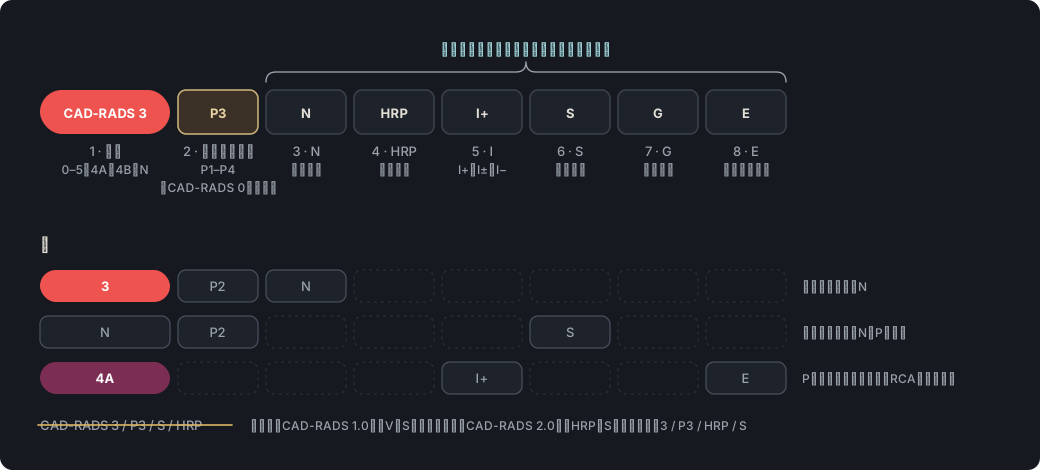
出典: CAD-RADS 2.0、表4、脚注 · “The CAD-RADS classification should be applied on a per-patient basis for the clinically most relevant (usually highest-grade) stenosis.”
CAD-RADS 0とCAD-RADS 1 CAD-RADS 0 versus CAD-RADS 1#
出典: CAD-RADS 2.0、表4、脚注b · “CAD-RADS 1 - This category should also include the presence of plaque with positive remodeling and no evidence of stenosis.”
出典: CAD-RADS 2.0、3.1.1節 · “In the presence of CAD-RADS 3, which reflects moderate stenosis (50–69%), there is an option to consider the use of CT-FFR, CTP, or stress testing”
出典: CAD-RADS 2.0、3.1.1節 · “It may be acute or chronic, and, in the context of chronic occlusion, factors such as lesion length, calcification particularly at the proximal aspect, tortuosity and degree of collateralization”
評価不能(N)のカテゴリーと修飾子 Nondiagnostic (N) category and modifierN#
別名:CAD-RADS N, N修飾子, 診断不能(nondiagnosticの直訳), nondiagnostic(英語)
出典: CAD-RADS 2.0、3.3.1節(Modifier N)、図12 · “for a patient with at least one non-interpretable segment and no stenosis (zero), minimal (1–24%), or no more than mild stenosis (25–49%) in interpretable segments, CAD-RADS N should be used”
CAD-RADSの記載順と修飾子 CAD-RADS coding order and modifiersP・N・HRP・I・S・G・E#
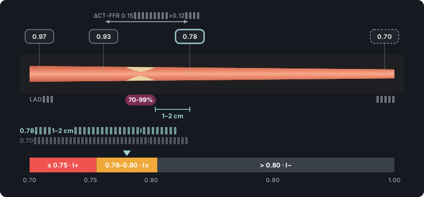
出典: CAD-RADS 2.0、3.3.5.1節、表6(Interpretation of CT-FFR) · “If there is presence of abnormal CT-FFR as defined as a lesion-specific value ≤ 0.75 in a vessel large enough for PCI the designation of “I+” (I=Ischemia) should be included.”
出典: CAD-RADS 2.0、3.3.5.2節、表7(Interpretation of Stress Myocardial CT Perfusion) · “If no ischemia is detected or if there is presence of a prior fixed myocardial infarct, the Modifier “I−” will be added to CAD-RADS.”
出典: CAD-RADS 2.0、1節、表4;SCCT 2026、表6、p. 52 · “the clinical management suggestions provided by CAD-RADS should not replace clinical judgment, particularly as there are many patient-specific factors that may influence clinical management.”
Rajiah PS, Alkadhi H, Andreini D, Bullock-Palmer RP, Chinnaiyan K, Chow B, et al. Interpretation and reporting of coronary computed tomographic angiography (2026 update): an expert consensus document of the Society of Cardiovascular Computed Tomography (SCCT). J Cardiovasc Comput Tomogr. 2026. In press. doi:10.1016/j.jcct.2026.08.014 Cited pages refer to the journal pre-proof PDF.
Cury RC, Leipsic J, Abbara S, Achenbach S, Berman D, Bittencourt M, et al. CAD-RADS 2.0 - 2022 Coronary Artery Disease-Reporting and Data System: an expert consensus document of the SCCT, ACC, ACR and NASCI. J Cardiovasc Comput Tomogr. 2022;16(6):536-57. doi:10.1016/j.jcct.2022.07.002
Koweek L, Achenbach S, Berman DS, Carr JJ, Cury RC, Ghoshhajra B, et al. Standardized medical terminology for cardiac computed tomography 2023 update: an expert consensus document of the SCCT, AAPM, ACR, NASCI and RSNA. J Cardiovasc Comput Tomogr. 2023;17(5):345-54. doi:10.1016/j.jcct.2023.06.002
Narula J, Chandrashekhar Y, Ahmadi A, Abbara S, Berman DS, Blankstein R, et al. SCCT 2021 expert consensus document on coronary computed tomographic angiography: a report of the Society of Cardiovascular Computed Tomography. J Cardiovasc Comput Tomogr. 2021;15(3):192-217. doi:10.1016/j.jcct.2020.11.001
Neves PO, Andrade J, Monção H. Coronary artery calcium score: current status. Radiol Bras. 2017;50(3):182-9.
Tamburino C, Tomasello SD, Capodanno D, Di Salvo ME, Marzà F, Galassi AR. Long-term follow-up after drug eluting stent implantation in left main trifurcations. EuroIntervention. 2009;5(4):432-7. doi:10.4244/eijv5i4a68
Kovacevic M, Burzotta F, Elharty S, Besis G, Aurigemma C, Romagnoli E, et al. Left main trifurcation and its percutaneous treatment: what is known so far? Circ Cardiovasc Interv. 2021;14(3):e009872. doi:10.1161/CIRCINTERVENTIONS.120.009872
Kim C, Park CH, Lee BY, et al. 2024 consensus statement on coronary stenosis and plaque evaluation in CT angiography from the Asian Society of Cardiovascular Imaging-Practical Tutorial (ASCI-PT). Korean J Radiol. 2024;25(4):331-42. doi:10.3348/kjr.2024.0112
「他の文献」で引用した文献
Sianos G, Morel MA, Kappetein AP, et al. The SYNTAX Score: an angiographic tool grading the complexity of coronary artery disease. EuroIntervention. 2005;1(2):219-27.
Cury RC, Abbara S, Achenbach S, et al. CAD-RADS: Coronary Artery Disease – Reporting and Data System. J Cardiovasc Comput Tomogr. 2016;10(4):269-81. doi:10.1016/j.jcct.2016.04.005
Williams MC, Moss AJ, Dweck M, et al. Coronary artery plaque characteristics associated with adverse outcomes in the SCOT-HEART study. J Am Coll Cardiol. 2019;73(3):291-301. doi:10.1016/j.jacc.2018.10.066
Hecht HS, Blaha MJ, Kazerooni EA, et al. CAC-DRS: Coronary Artery Calcium Data and Reporting System. An expert consensus document of the Society of Cardiovascular Computed Tomography (SCCT). J Cardiovasc Comput Tomogr. 2018;12(3):185-91. doi:10.1016/j.jcct.2018.03.008
Lawton JS, Tamis-Holland JE, Bangalore S, et al. 2021 ACC/AHA/SCAI guideline for coronary artery revascularization: a report of the American College of Cardiology/American Heart Association Joint Committee on Clinical Practice Guidelines. J Am Coll Cardiol. 2022;79(2):e21-e129. doi:10.1016/j.jacc.2021.09.006
Galassi AR, Werner GS, Boukhris M, et al. Percutaneous recanalisation of chronic total occlusions: 2019 consensus document from the EuroCTO Club. EuroIntervention. 2019;15(2):198-208. doi:10.4244/EIJ-D-18-00826
Tonino PA, De Bruyne B, Pijls NH, et al. Fractional flow reserve versus angiography for guiding percutaneous coronary intervention. N Engl J Med. 2009;360(3):213-24. doi:10.1056/NEJMoa0807611
Gulati M, Levy PD, Mukherjee D, et al. 2021 AHA/ACC/ASE/CHEST/SAEM/SCCT/SCMR Guideline for the Evaluation and Diagnosis of Chest Pain: A Report of the American College of Cardiology/American Heart Association Joint Committee on Clinical Practice Guidelines. Circulation. 2021;144(22):e368-e454. doi:10.1161/CIR.0000000000001029