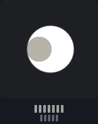
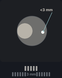
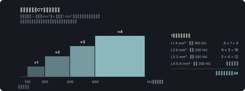
出典: SCCT 2026、動脈硬化性プラークの項、p. 13 · “(largely) calcified plaque: attenuation values of most of the plaque exceed the values of the contrast-enhanced blood”
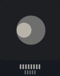
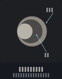
出典: SCCT 2026、動脈硬化性プラークの項、p. 13 · “partially calcified: with both calcified and non-calcified components (Fig 6). A partially calcified plaque can be specified as predominantly calcified with ≥ 75 % of the plaque component calcified”
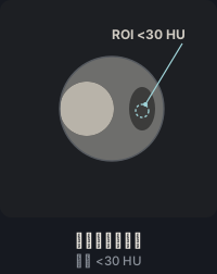
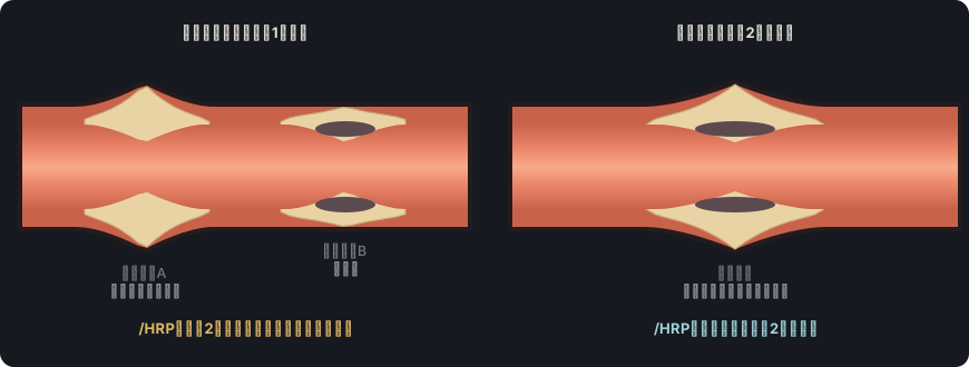
出典: CAD-RADS 2.0、3.3.4節、図15 · “If a coronary plaque clearly demonstrates two or more high-risk features by CCTA, the modifier “HRP” (high risk plaque) should be added”
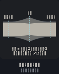
出典: CAD-RADS 2.0、図15;SCCT 2026、p. 13 · “Positive remodeling, defined as the ratio of outer vessel diameter at the site of plaque divided by the average outer diameter of the proximal and distal vessel greater than 1.1”
出典: SCCT 2026、本文および表3、pp. 14・49;CAD-RADS 2.0、3.2.1節 · “CAD-RADS 2.0 introduces the P1 to P4 descriptors which are used to denote increasing categories of plaque burden (P1=mild, P2=moderate, P3=severe and P4=extensive)”
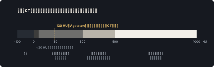
出典: Neves PO, et al. Radiol Bras 2017(総説)、「Agatston method」の項 · “The Agatston method uses the weighted sum of lesions with a density above 130 HU, multiplying the area of calcium by a factor related to maximum plaque attenuation”
Rajiah PS, Alkadhi H, Andreini D, Bullock-Palmer RP, Chinnaiyan K, Chow B, et al. Interpretation and reporting of coronary computed tomographic angiography (2026 update): an expert consensus document of the Society of Cardiovascular Computed Tomography (SCCT). J Cardiovasc Comput Tomogr. 2026. In press. doi:10.1016/j.jcct.2026.08.014 Cited pages refer to the journal pre-proof PDF.
Cury RC, Leipsic J, Abbara S, Achenbach S, Berman D, Bittencourt M, et al. CAD-RADS 2.0 - 2022 Coronary Artery Disease-Reporting and Data System: an expert consensus document of the SCCT, ACC, ACR and NASCI. J Cardiovasc Comput Tomogr. 2022;16(6):536-57. doi:10.1016/j.jcct.2022.07.002
Koweek L, Achenbach S, Berman DS, Carr JJ, Cury RC, Ghoshhajra B, et al. Standardized medical terminology for cardiac computed tomography 2023 update: an expert consensus document of the SCCT, AAPM, ACR, NASCI and RSNA. J Cardiovasc Comput Tomogr. 2023;17(5):345-54. doi:10.1016/j.jcct.2023.06.002
Narula J, Chandrashekhar Y, Ahmadi A, Abbara S, Berman DS, Blankstein R, et al. SCCT 2021 expert consensus document on coronary computed tomographic angiography: a report of the Society of Cardiovascular Computed Tomography. J Cardiovasc Comput Tomogr. 2021;15(3):192-217. doi:10.1016/j.jcct.2020.11.001
Neves PO, Andrade J, Monção H. Coronary artery calcium score: current status. Radiol Bras. 2017;50(3):182-9.
Tamburino C, Tomasello SD, Capodanno D, Di Salvo ME, Marzà F, Galassi AR. Long-term follow-up after drug eluting stent implantation in left main trifurcations. EuroIntervention. 2009;5(4):432-7. doi:10.4244/eijv5i4a68
Kovacevic M, Burzotta F, Elharty S, Besis G, Aurigemma C, Romagnoli E, et al. Left main trifurcation and its percutaneous treatment: what is known so far? Circ Cardiovasc Interv. 2021;14(3):e009872. doi:10.1161/CIRCINTERVENTIONS.120.009872
Kim C, Park CH, Lee BY, et al. 2024 consensus statement on coronary stenosis and plaque evaluation in CT angiography from the Asian Society of Cardiovascular Imaging-Practical Tutorial (ASCI-PT). Korean J Radiol. 2024;25(4):331-42. doi:10.3348/kjr.2024.0112
「他の文献」で引用した文献
Leipsic J, Abbara S, Achenbach S, Cury R, Earls JP, Mancini GBJ, et al. SCCT guidelines for the interpretation and reporting of coronary CT angiography: a report of the Society of Cardiovascular Computed Tomography Guidelines Committee. J Cardiovasc Comput Tomogr. 2014;8(5):342-58. doi:10.1016/j.jcct.2014.07.003
Cury RC, Abbara S, Achenbach S, et al. CAD-RADS: Coronary Artery Disease – Reporting and Data System. J Cardiovasc Comput Tomogr. 2016;10(4):269-81. doi:10.1016/j.jcct.2016.04.005
Nieman K, García-García HM, Hideo-Kajita A, et al. Standards for quantitative assessments by coronary computed tomography angiography (CCTA): an expert consensus document of the SCCT. J Cardiovasc Comput Tomogr. 2024;18(5):429-43. doi:10.1016/j.jcct.2024.05.232
Williams MC, Moss AJ, Dweck M, et al. Coronary artery plaque characteristics associated with adverse outcomes in the SCOT-HEART study. J Am Coll Cardiol. 2019;73(3):291-301. doi:10.1016/j.jacc.2018.10.066
van Rosendael SE, Shiyovich A, Cardoso RN, Souza Freire CV, van Rosendael AR, Lin FY, et al. The role of cardiac computed tomography angiography in risk stratification for coronary artery disease. J Soc Cardiovasc Angiogr Interv. 2024;3(11):102230. doi:10.1016/j.jscai.2024.102230
Yang S, Lee JM, Hoshino M, Murai T, Choi KH, Hwang D, et al. Prognostic implications of comprehensive whole vessel plaque quantification using coronary computed tomography angiography. JACC Asia. 2021;1(1):37-48. doi:10.1016/j.jacasi.2021.05.003
Pozo E, Agudo-Quilez P, Rojas-González A, Alvarado T, Olivera MJ, Jiménez-Borreguero LJ, et al. Noninvasive diagnosis of vulnerable coronary plaque. World J Cardiol. 2016;8(9):520-33. doi:10.4330/wjc.v8.i9.520
Bittencourt MS, Hulten E, Ghoshhajra B, et al. Prognostic value of nonobstructive and obstructive coronary artery disease detected by coronary computed tomography angiography to identify cardiovascular events. Circ Cardiovasc Imaging. 2014;7(2):282-91. doi:10.1161/CIRCIMAGING.113.001047
Hecht HS, Blaha MJ, Kazerooni EA, et al. CAC-DRS: Coronary Artery Calcium Data and Reporting System. An expert consensus document of the Society of Cardiovascular Computed Tomography (SCCT). J Cardiovasc Comput Tomogr. 2018;12(3):185-91. doi:10.1016/j.jcct.2018.03.008
Obisesan OH, Osei AD, Uddin SMI, Dzaye O, Blaha MJ. An update on coronary artery calcium interpretation at chest and cardiac CT. Radiol Cardiothorac Imaging. 2021;3(1):e200484. doi:10.1148/ryct.2021200484