cctamap.comTutorial
CCTA MAP · TUTORIAL 01

Your first CAD-RADS map

One case, seven steps.

From the coronary CTA findings to the map, the category and the draft report. Anonymized teaching case · about 3 minutes.

The case

  • Right dominance, no ramus intermedius.
  • Partially calcified plaque in the proximal LAD, at the origin of the first diagonal.
  • Severe stenosis (70–99%).
  • Positive remodeling and napkin-ring sign.
  • Calcium score (Agatston): 29.

When you finish, the map will read CAD-RADS 4A/P1/HRP

The case on coronary CTA

Before opening the map, this is what the study shows.

Axial. Scrolling through the slices, a plaque appears in the proximal LAD.
Curved MPR of the LAD. The plaque narrows the lumen: severe stenosis (70–99%).
Cross-sections. A low-attenuation core with a denser rim, the napkin-ring sign, and a vessel wall that bulges outward, positive remodeling. Bottom row: proximal reference, plaque and distal reference.
Q, MDOne lesion, several findings: grade, plaque type, high-risk features and calcium. All of that goes on the map.
  1. Dominance
  2. Draw the lesion
  3. Grade and plaque
  4. Plaque burden
  5. Category
  6. Report
  7. Share the case

Step 1

Make the tree match the patient's

Before marking lesions, choose the dominance (right, left or balanced) and say whether there is a ramus intermedius. The map is redrawn: it changes which artery gives rise to the posterior descending artery, and the ramus appears or disappears.

In this case: Right, with the ramus intermedius switched off.

ClaudiaOn a phone these buttons are below the map. On a computer, right next to it.
Each dominance redraws the tree. The case ends up right dominant, with no ramus intermedius.

Step 2

Draw the lesion on the vessel

Rest your finger on the vessel for a moment, then slide it along the plaque; with a mouse, drag. The lesion is drawn exactly over that stretch.

If you just tap, without dragging, the editor opens for the whole segment.
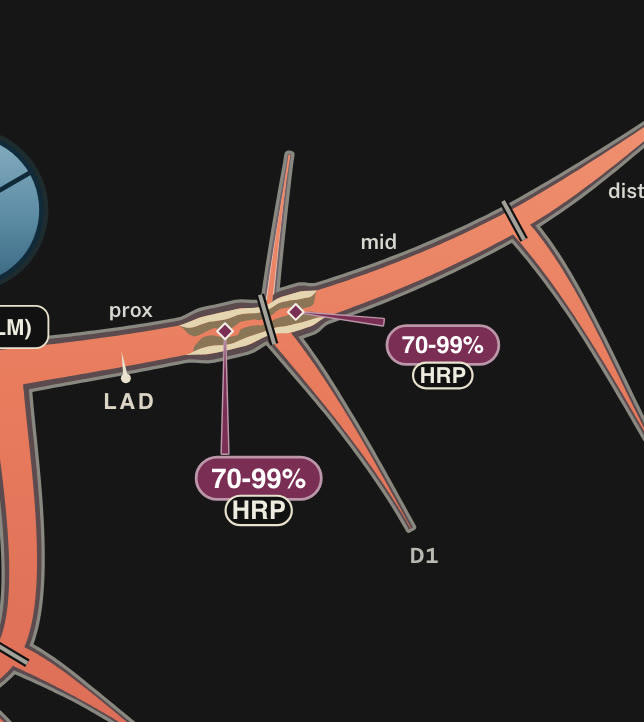
Close-up, already graded: the lesion crosses the segment boundary (the dark bar), where the first diagonal arises.
Q, MDWhat if the lesion sits right on the boundary between two segments?
ClaudiaIt belongs to the preceding segment. Cross the boundary only when the plaque truly continues into the next one; then the SIS counts both.SCCT 2026 convention, section 2.2.1.
Rest, slide and release: the lesion editor opens.

Step 3

Grade, plaque type and high-risk features

When you release, the lesion editor opens. Choose:

  • Severity: 4 sev, a 70–99% stenosis. If you measured the percentage, set it with the slider.
  • Plaque type: Partly calc.
  • High-risk features: Positive remodeling and Napkin-ring sign.

At the top you see the segments the lesion spans. You can give each one a different grade, or remove one with ✕.

ClaudiaWith two features in the same plaque, the counter reads HRP 2/4 and the plaque is flagged as high-risk.
Grade 4, partially calcified plaque and two high-risk features.

Step 4

Plaque burden: the P modifier

Open Modifiers. Plaque burden can be obtained in three ways:

  • Auto (map): counts the segments with plaque (SIS).
  • Calcium (Agatston): you enter the calcium score.
  • Pick P: you choose it yourself, for instance by visual assessment.

Here there is a calcium score: Calcium (Agatston) and 29. Between 1 and 100 corresponds to P1.

The same panel holds ischemia (the I modifier), which only appears if you record a functional test.

Q, MDNo calcium score? I leave “Auto (map)” on and P comes from the number of segments with plaque.
Agatston 29: mild plaque burden, P1.

Step 5

The category is calculated for you

With every change, the map updates the category and its modifiers. For this case: CAD-RADS 4A/P1/HRP

4ASevere stenosis (70–99%) in one or two vessels. Here, the LAD.
P1Mild plaque burden: Agatston 29.
HRPHigh-risk plaque: two features in the same plaque, positive remodeling and napkin-ring sign.

The Report tab shows this same breakdown with your data.

Claudia4A/P1/HRP! And “Animate flow” shows the blood flowing through the vessels.
The lesion takes the color of its grade; the category sits at the top.

Step 6

Copy the draft report

The Report tab assembles the findings, the impression and the management suggested by the consensus. Copy it, or download it as Word or PDF.

FINDINGS

Left anterior descending (LAD): partially calcified plaque extending from the proximal to the mid segment, causing severe stenosis (70-99%), with high-risk plaque features (positive remodeling and napkin-ring sign); the remainder is unremarkable. Branches D1, D2: no plaque.

IMPRESSION

Severe obstructive CAD. CAD-RADS 4A/P1/HRP.

Anything you did not mark is described as normal.

Q, MDIt is a draft. I check it against the study before signing.
The category, the reasons for it and, further down, the draft.

Step 7

Save or share the case

Case link / QR gives you a link and a QR code that reopen the map exactly as you left it. Map image downloads the tree for your report.

ClaudiaThe case travels inside the link. It is not sent to any server; only anonymous usage statistics are counted.

Open this case in the map

Case link and QR code.

On a computer it works the same

On a large screen the report sits beside the map, and the case buttons right next to it. The steps are the same, with click and drag.
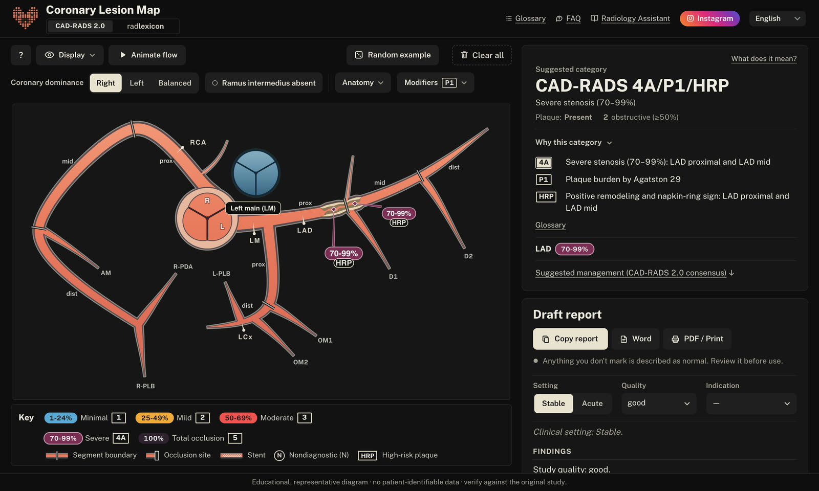
The same case in the computer view

Keep going