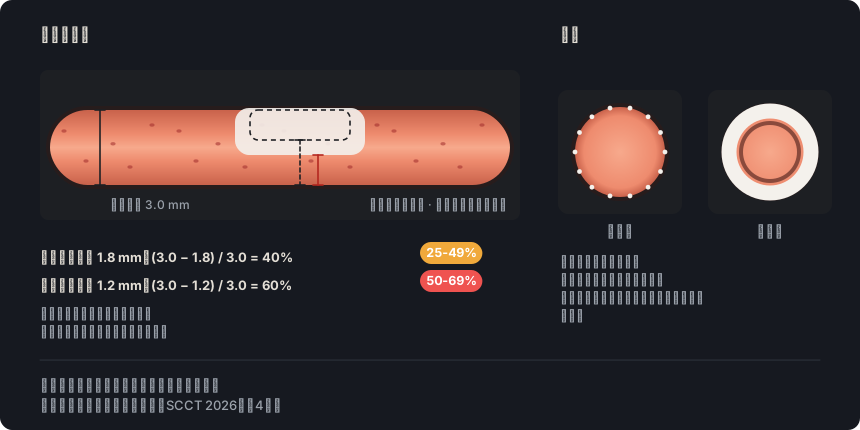
出处: SCCT 2026,报告部分,d 项“Image quality and findings”,及表6,第35、52页 · “With no established standards for overall study quality, a visual schema such as excellent, good, average, fair, poor or non-diagnostic is recommended.”
出处: SCCT 2026,报告部分,d 项“Image quality and findings”;另见狭窄部分,第15页 · “Whenever coronary segments are not interpretable, the affected segments and the causative artifact should be stated.”
详情
实际应用: 判定无法评估之前,先在其他时相、其他平面和其他窗宽窗位下复核。若仍无法判读,写明节段和伪影,并描述可见的斑块。存在 N 时,CAD-RADS 2.0 仅在整体斑块负荷可以可靠评估时(例如借助钙化积分)才加上 P。其对分类的影响见 CAD-RADS 一节。
出处: SCCT 2026,Image quality, artifacts & pitfalls,c 项 Motion artifacts,第25–26页 · “Cardiac motion appears as vessel blurring, most commonly in the highly mobile vertical segment of the right coronary artery”
出处: SCCT 2026,Image quality, artifacts & pitfalls,c 项 Motion artifacts,第26页 · “Transition (stairstep or misalignment) artifacts occur at slab interfaces from different cardiac cycles, typically due to irregular rhythm or incorrect rhythm gating”
出处: SCCT 2026,Image quality, artifacts & pitfalls,e 项 Blooming artifact;表5,第27、51页 · “Blooming artifact results from partial volume averaging within a voxel, causing blurring at interfaces between coronary arteries and calcified plaques or stents and leading to overestimation of luminal stenosis”
出处: SCCT 2026,Image quality, artifacts & pitfalls,d 项 Beam hardening and metallic artifacts;心肌部分,第27、31页 · “Beam-hardening artifacts arise from preferential attenuation of low-energy photons by metal, calcium, or dense contrast, producing low-attenuation areas or streaks adjacent to bright artifacts”
出处: SCCT 2026,Image quality, artifacts & pitfalls,a 项 Excessive noise;表4,第25、50页 · “Image noise from low photon counts appears as granular texture with random bright and dark streaks.”
出处: SCCT 2026,图像格式,b 项 Multiplanar reformats 和 c 项 Curved and straightened MPR,第8–9页;冠状动脉判读,第11页;动脉粥样硬化斑块,第13页;狭窄的定性与定量分级,第15–16页;假阴性、假阳性与高估,第28–29页 · “Attention should be paid to post-processing artifacts including centerline misregistration, which can result in false-positive stenosis.”
出处: SCCT 2026,图像格式,d 项 Maximal intensity projection 和 h 项 Other image types,第9、11页;假阴性和假阳性,第28页;心肌,第30–31页。SCCT 2023 术语,表3 · “MIP should not be interpreted in isolation because it lacks in-depth attenuation information and is unreliable for stenosis assessment, underestimating noncalcified plaque and overestimating calcified disease”
Rajiah PS, Alkadhi H, Andreini D, Bullock-Palmer RP, Chinnaiyan K, Chow B, et al. Interpretation and reporting of coronary computed tomographic angiography (2026 update): an expert consensus document of the Society of Cardiovascular Computed Tomography (SCCT). J Cardiovasc Comput Tomogr. 2026. 印刷中。 doi:10.1016/j.jcct.2026.08.014 所引页码对应预印本PDF(journal pre-proof)。
Cury RC, Leipsic J, Abbara S, Achenbach S, Berman D, Bittencourt M, et al. CAD-RADS 2.0 - 2022 Coronary Artery Disease-Reporting and Data System: an expert consensus document of the SCCT, ACC, ACR and NASCI. J Cardiovasc Comput Tomogr. 2022;16(6):536-57. doi:10.1016/j.jcct.2022.07.002
Koweek L, Achenbach S, Berman DS, Carr JJ, Cury RC, Ghoshhajra B, et al. Standardized medical terminology for cardiac computed tomography 2023 update: an expert consensus document of the SCCT, AAPM, ACR, NASCI and RSNA. J Cardiovasc Comput Tomogr. 2023;17(5):345-54. doi:10.1016/j.jcct.2023.06.002
Narula J, Chandrashekhar Y, Ahmadi A, Abbara S, Berman DS, Blankstein R, et al. SCCT 2021 expert consensus document on coronary computed tomographic angiography: a report of the Society of Cardiovascular Computed Tomography. J Cardiovasc Comput Tomogr. 2021;15(3):192-217. doi:10.1016/j.jcct.2020.11.001
Neves PO, Andrade J, Monção H. Coronary artery calcium score: current status. Radiol Bras. 2017;50(3):182-9.
Tamburino C, Tomasello SD, Capodanno D, Di Salvo ME, Marzà F, Galassi AR. Long-term follow-up after drug eluting stent implantation in left main trifurcations. EuroIntervention. 2009;5(4):432-7. doi:10.4244/eijv5i4a68
Kovacevic M, Burzotta F, Elharty S, Besis G, Aurigemma C, Romagnoli E, et al. Left main trifurcation and its percutaneous treatment: what is known so far? Circ Cardiovasc Interv. 2021;14(3):e009872. doi:10.1161/CIRCINTERVENTIONS.120.009872
Kim C, Park CH, Lee BY, et al. 2024 consensus statement on coronary stenosis and plaque evaluation in CT angiography from the Asian Society of Cardiovascular Imaging-Practical Tutorial (ASCI-PT). Korean J Radiol. 2024;25(4):331-42. doi:10.3348/kjr.2024.0112
“其他文献”注释的出处
Leipsic J, Abbara S, Achenbach S, Cury R, Earls JP, Mancini GBJ, et al. SCCT guidelines for the interpretation and reporting of coronary CT angiography: a report of the Society of Cardiovascular Computed Tomography Guidelines Committee. J Cardiovasc Comput Tomogr. 2014;8(5):342-58. doi:10.1016/j.jcct.2014.07.003
Cury RC, Abbara S, Achenbach S, et al. CAD-RADS: Coronary Artery Disease – Reporting and Data System. J Cardiovasc Comput Tomogr. 2016;10(4):269-81. doi:10.1016/j.jcct.2016.04.005
Nieman K, García-García HM, Hideo-Kajita A, et al. Standards for quantitative assessments by coronary computed tomography angiography (CCTA): an expert consensus document of the SCCT. J Cardiovasc Comput Tomogr. 2024;18(5):429-43. doi:10.1016/j.jcct.2024.05.232
Koweek L, Achenbach S, Berman DS, et al. Standardized medical terminology for cardiac computed tomography 2023 update: an expert consensus document of the Society of Cardiovascular Computed Tomography (SCCT), American Association of Physicists in Medicine (AAPM), American College of Radiology (ACR), North American Society for Cardiovascular Imaging (NASCI), and Radiological Society of North America (RSNA). Radiol Cardiothorac Imaging. 2023;5(4):e230167. doi:10.1148/ryct.230167
Abbara S, Blanke P, Maroules CD, et al. SCCT guidelines for the performance and acquisition of coronary computed tomographic angiography: a report of the Society of Cardiovascular Computed Tomography Guidelines Committee. Endorsed by the North American Society for Cardiovascular Imaging (NASCI). J Cardiovasc Comput Tomogr. 2016;10(6):435-49. doi:10.1016/j.jcct.2016.10.002