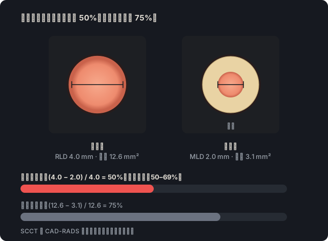
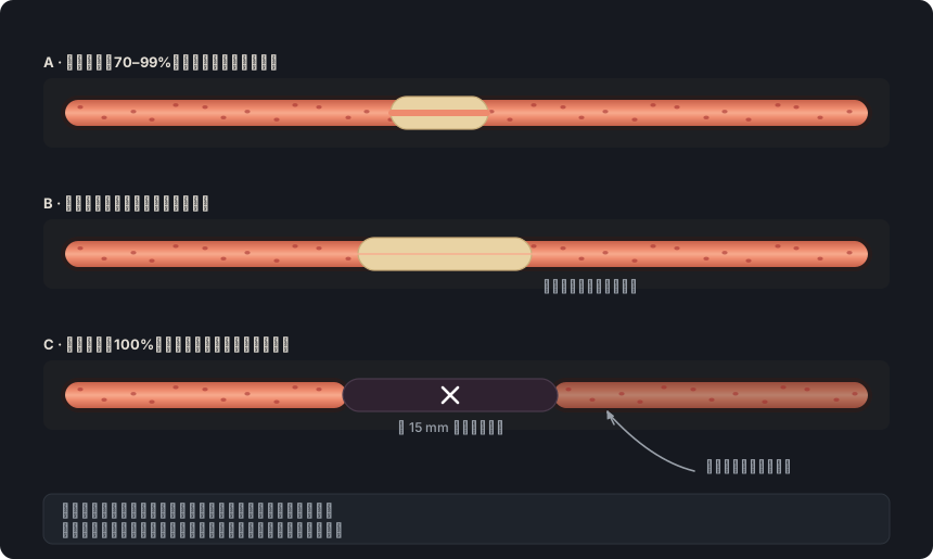
出处: SCCT 2026,狭窄定性分级正文,p. 15;曲面重组,p. 9;定量分级中的公式,p. 16 · “A qualitative interpretation of the extent of coronary artery stenosis should be made in transverse and longitudinal views as an estimated maximal percent diameter stenosis.”
出处: SCCT 2026,狭窄定性分级(p. 15)与定量分级(pp. 16–17)正文;高估,p. 29 · “Qualitatively, the luminal diameter at a stenosis should be compared with the luminal diameter of a normal reference vessel segment distal and/or proximal to the stenosis.”
出处: SCCT 2026,狭窄定性分级正文,p. 15 · “This stenosis can be categorized as minimal (1-24 %), mild (25-49 %), moderate (50-69 %), severe (70-99 %) and total occlusion (100 %)”
出处: SCCT 2026,冠状动脉支架(p. 19)、桥血管(pp. 19–20)、动脉粥样硬化斑块(p. 13)及印象(p. 36)正文;CAD-RADS 2.0,表1(0%,“No visible stenosis”)及表4(CAD-RADS 0和1,注b) · “An impression of a normal study should be given only if there is no plaque or stenosis.”
出处: CAD-RADS 2.0,第3.1.1节及表4(CAD-RADS 1和2为非阻塞性病变;CAD-RADS 3至4B的解读) · “CAD RADS 4B - This indicates the presence of a left main stenosis of at least 50% or three-vessel obstructive disease (>70%).”
出处: SCCT 2026,动脉粥样硬化斑块与斑块体积正文(p. 14)及表6(按节段评估斑块与狭窄),p. 52;CAD-RADS 2.0,第3.1.1和3.3.4节、表4(按患者分类的注释)及图17 · “Depending on the extent of disease, plaque can be described on a per-lesion or per-segment level.”
出处: SCCT 2026,动脉粥样硬化斑块(p. 14)、定量分级(pp. 16–17)及低估(p. 29)正文 · “Additional attributes such as "ostial", "bifurcation", "eccentric", "long", "diffuse" and "multifocal" can be used to further describe the location, morphology and extent of atherosclerotic disease.”
出处: SCCT 2026,动脉粥样硬化斑块(“eccentric”属性,p. 14)、曲面重组(p. 9)及血管炎(p. 24)正文;CAD-RADS 2.0,第3.3.6节;Kim 2024(ASCI-PT),主题8 · “Curved MPR can underestimate eccentric stenosis; hence evaluation should be performed in at least two perpendicular planes.”
出处: SCCT 2026,狭窄定量分级正文,p. 17 · “Visual estimate is typically sufficient for clinical interpretation since it is comparable to visual stenosis assessment in ICA”
出处: SCCT 2026,表5(判读共识,强推荐)及表6(按CAD-RADS 2.0逐节段评估);书写示例,p. 15 · “Description of stenosis is based on the anatomical location (e.g., mid LAD, distal RCA) rather than on segment numbers (e.g. segment 5, segment 7).”
出处: SCCT 2026,动脉粥样硬化斑块(“ostial”属性,p. 14)、冠状动脉解剖(p. 11)、定量分级(p. 16)、MIP(p. 9)、起源异常(p. 20)及血管炎(p. 24)正文;Kim 2024(ASCI-PT),主题1和7;CAD-RADS 2.0,表4及第3.3.6节 · “If the proximal segment is not appropriate (e.g., ostial lesion), the closest distal reference can be chosen.”
Rajiah PS, Alkadhi H, Andreini D, Bullock-Palmer RP, Chinnaiyan K, Chow B, et al. Interpretation and reporting of coronary computed tomographic angiography (2026 update): an expert consensus document of the Society of Cardiovascular Computed Tomography (SCCT). J Cardiovasc Comput Tomogr. 2026. 印刷中。 doi:10.1016/j.jcct.2026.08.014 所引页码对应预印本PDF(journal pre-proof)。
Cury RC, Leipsic J, Abbara S, Achenbach S, Berman D, Bittencourt M, et al. CAD-RADS 2.0 - 2022 Coronary Artery Disease-Reporting and Data System: an expert consensus document of the SCCT, ACC, ACR and NASCI. J Cardiovasc Comput Tomogr. 2022;16(6):536-57. doi:10.1016/j.jcct.2022.07.002
Koweek L, Achenbach S, Berman DS, Carr JJ, Cury RC, Ghoshhajra B, et al. Standardized medical terminology for cardiac computed tomography 2023 update: an expert consensus document of the SCCT, AAPM, ACR, NASCI and RSNA. J Cardiovasc Comput Tomogr. 2023;17(5):345-54. doi:10.1016/j.jcct.2023.06.002
Narula J, Chandrashekhar Y, Ahmadi A, Abbara S, Berman DS, Blankstein R, et al. SCCT 2021 expert consensus document on coronary computed tomographic angiography: a report of the Society of Cardiovascular Computed Tomography. J Cardiovasc Comput Tomogr. 2021;15(3):192-217. doi:10.1016/j.jcct.2020.11.001
Neves PO, Andrade J, Monção H. Coronary artery calcium score: current status. Radiol Bras. 2017;50(3):182-9.
Tamburino C, Tomasello SD, Capodanno D, Di Salvo ME, Marzà F, Galassi AR. Long-term follow-up after drug eluting stent implantation in left main trifurcations. EuroIntervention. 2009;5(4):432-7. doi:10.4244/eijv5i4a68
Kovacevic M, Burzotta F, Elharty S, Besis G, Aurigemma C, Romagnoli E, et al. Left main trifurcation and its percutaneous treatment: what is known so far? Circ Cardiovasc Interv. 2021;14(3):e009872. doi:10.1161/CIRCINTERVENTIONS.120.009872
Kim C, Park CH, Lee BY, et al. 2024 consensus statement on coronary stenosis and plaque evaluation in CT angiography from the Asian Society of Cardiovascular Imaging-Practical Tutorial (ASCI-PT). Korean J Radiol. 2024;25(4):331-42. doi:10.3348/kjr.2024.0112
“其他文献”注释的出处
Sianos G, Morel MA, Kappetein AP, et al. The SYNTAX Score: an angiographic tool grading the complexity of coronary artery disease. EuroIntervention. 2005;1(2):219-27.
Leipsic J, Abbara S, Achenbach S, Cury R, Earls JP, Mancini GBJ, et al. SCCT guidelines for the interpretation and reporting of coronary CT angiography: a report of the Society of Cardiovascular Computed Tomography Guidelines Committee. J Cardiovasc Comput Tomogr. 2014;8(5):342-58. doi:10.1016/j.jcct.2014.07.003
Nieman K, García-García HM, Hideo-Kajita A, et al. Standards for quantitative assessments by coronary computed tomography angiography (CCTA): an expert consensus document of the SCCT. J Cardiovasc Comput Tomogr. 2024;18(5):429-43. doi:10.1016/j.jcct.2024.05.232
Raff GL, Abidov A, Achenbach S, et al. SCCT guidelines for the interpretation and reporting of coronary computed tomographic angiography. J Cardiovasc Comput Tomogr. 2009;3(2):122-36. doi:10.1016/j.jcct.2009.01.001
Galassi AR, Vadalà G, Werner GS, et al. Evaluation and management of patients with coronary chronic total occlusions considered for revascularisation. A clinical consensus statement of the European Association of Percutaneous Cardiovascular Interventions (EAPCI) of the ESC, the European Association of Cardiovascular Imaging (EACVI) of the ESC, and the ESC Working Group on Cardiovascular Surgery. EuroIntervention. 2024;20(3):e174-84. doi:10.4244/EIJ-D-23-00749
Lawton JS, Tamis-Holland JE, Bangalore S, et al. 2021 ACC/AHA/SCAI guideline for coronary artery revascularization: a report of the American College of Cardiology/American Heart Association Joint Committee on Clinical Practice Guidelines. J Am Coll Cardiol. 2022;79(2):e21-e129. doi:10.1016/j.jacc.2021.09.006